Introduction
Magnetic Resonance Imaging (MRI) has revolutionized the field of neuroimaging by offering unparalleled comprehensions into the structural and functional characteristics of the human brain. In recent years, MRI-based morphometric analysis has become a powerful tool for investigating the anatomical variations and dimensions of specific brain regions, providing valuable information for understanding neurodevelopmental processes, assessing pathological conditions, and even predicting cognitive functions.1 In MRI studies of brain structures, particular focus has been directed towards the corpus callosum (CC), a significant white matter tract crucial for interhemispheric communication. It plays a pivotal role in integrating sensory, motor, and cognitive information across cerebral hemispheres, facilitating complex cognitive functions. 2, 3 The CC, conventionally divided into regions like the rostrum, genu, body, and splenium, establishes somatotropic connections with specific cortical areas, enabling efficient neural communication. Research indicates that CC dimensions exhibit variability influenced by factors such as age, sex, racial background, and various pathological conditions. 4, 5 In a seminal study by Witelson, it was observed that the CC tends to be larger in individuals who are left-handed or ambidextrous, potentially reflecting reduced cortical specialization and an increased need for interhemispheric communication. 6 Additionally, investigations have highlighted a larger splenial dimension in women compared to men, hypothesized to relate to superior auditory and visual processing abilities in women, controlled by the temporal and occipital lobes, respectively, and fused across the midline by splenial fibers. 6, 7, 8 However, conflicting evidence exists regarding the presence of this sexual dimorphism. 9, 10 Notably, while it is commonly presumed that CC dimensions decrease with age in tandem with brain atrophy, some studies have failed to corroborate this assertion.11 Research exploring the relationship between anatomical dimensions of the Corpus callosum (CC), information integration, and various diseases has captured the interest of neuroscientists and radiologists alike. A recent meta-analysis underscored a consistent observation of diminished dimensions of the CC in individuals diagnosed with schizophrenia and bipolar disorders. 12 This reduction was linked to a decline in white matter volume within the CC, resulting in impaired interhemispheric communication. Similarly, Khasawneh et al., in a recent investigation, reported a significant diminution in CC size among patients diagnosed with Alzheimer’s disease compared to control subjects.13 While extensive research has been conducted on corpus callosum dimensions in diverse populations worldwide, there remains a paucity of data concerning specific ethnic or regional groups. This gap in knowledge is particularly evident in regions such as Kashmir, where unique genetic, environmental, and sociocultural factors may influence brain anatomy and development. Therefore, this study aims to address this gap by characterizing the normative range and variability of corpus callosum dimensions in this population, we seek to contribute to a deeper understanding of brain morphology in a region characterized by distinct genetic and environmental influences. Furthermore, this research may have implications for clinical practice, such as improving diagnostic accuracy and treatment planning for neurological and psychiatric disorders prevalent in the Kashmiri population.
Materials and Methods
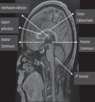
The present study was conducted within the premises of the Department of Anatomy, GMC Srinagar, spanning a duration of one year. A cohort of 300 patients was selected randomly for the study, drawing subjects from individuals who frequented the Hospital for routine brain MRI examinations. “During the study duration, a total of 5,921 images were meticulously reviewed, out of which 300 images met the stringent inclusion criteria and were consequently enrolled in the investigation. The acquired neuroimaging data underwent meticulous scrutiny by two independent Neuroradiologists, with a primary focus on discerning scans exhibiting normal anatomical features. Subjects presenting pathological findings, including neoplasms, infections, traumatic lesions, hydrocephalus, demyelinating abnormalities, and congenital malformations, were systematically excluded from the study cohort. Ethical approval for the research was duly obtained from the institutional ethics committee. The imaging procedures were conducted using a state-of-the-art 1.5T GE Healthcare MRI scanner equipped with a 16-channel head coil. The acquisition protocol encompassed Fast Spin Echo (FSE) T1-weighted imaging (T1WI), T2-weighted imaging (T2WI), and Fluid-Attenuated Inversion Recovery (FLAIR) sequences in axial, coronal, and sagittal planes. Specific imaging parameters included a repetition time (TR) of 744 milliseconds, an echo time (TE) of 2.47 milliseconds, a slice thickness of 1.60 millimeters, and a number of excitations (NEX) set at 1.0. Diligent radiographers adhered consistently to the established acquisition protocol. The resulting neuroimages were systematically archived within the K-PACS workstation version 1.5, alongside the image retrieval systems. Subsequently, a precise assessment of the corpus callosum (CC) dimensions was performed in the mid-sagittal plane. The CC measurements were determined based on the midpoints of the anterior and posterior commissures, as well as the interhemispheric fissure. This rigorous approach, in alignment with methodologies described by Allouh et al. and Pettey and Gee, ensured that only subtle cortical traces were visible on either side (Figure 1). 14 Additionally, the forebrain length (LM) was quantified by measuring the fronto-occipital diameter in the midsagittal plane at the midline."
Figure 1
Showcases a mid-sagittal magnetic resonance imaging (MRI) scan of the brain, delineating the corpus callosum.

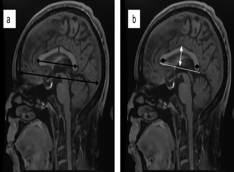
Figure 2
a: Elucidates the anterior-posterior extent of the corpus callosum and the fronto-occipital dimension of the brain, denoted by dual-ended arrows in black; b: The vertical dimension of the corpus callosum is depicted with a white dual-ended arrow, while the thickness of the genu and splenium is highlighted by black diamond-shaped arrows.

The evaluation of the corpus callosum (CC) entailed quantifying its anterior-posterior diameter (AP) from the foremost to the rearmost point within the mid-sagittal plane. Furthermore, the vertical dimension was assessed from the utmost superior to the utmost inferior point of the CC, as illustrated in (Figure 2a,b). These detailed measurements formed the foundation for subsequent analysis. The corpus callosum (CC) was subjected to segmentation utilizing specialized computer measurement software, adhering to a methodology originally advocated by Witelson, albeit with specific adaptations implemented across diverse morphometric studies of corpus callosa. To demarcate its subregions, five perpendicular lines were strategically placed along the maximal extension of the CC, yielding seven discernible subregions: the rostrum, genu, anterior body, mid-body, posterior body, isthmus, and splenium, as illustrated in (Figure 3a). Subsequently, the maximal diameters of these segments were measured with precision (Figure 3b).
Statistical analysis
Statistical analysis, performed using SPSS version 22, included a one-way analysis of variance (ANOVA) test to discern variations in mean CC dimensions among distinct age groups. Pearson’s correlation analysis elucidated the relationship between age and CC dimensions. The independent t-test compared mean values and variabilities of CC dimensions, considering the subjects' sex.
Results
In this section, the results of the study will be described:
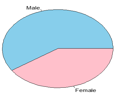
The cohort demonstrated a mean age of 46.01 ± 19.02 years, characterized by a male predominance represented by a ratio of 1.45:1. Evaluation of the average fronto-occipital diameter in the mid-sagittal plane revealed measurements of 163.21 ± 19.80 mm for males, while 159.08 ± 22.8 mm for females. However, statistical analysis indicated no significant difference between genders (p = 0.08).(Figure 4)
Table 1
Quantitativeanalysis of corpus callosum dimensions in adult individuals: A morphometric study
|
Dimensions |
Mean±SD |
|
Length(AB) |
74.815±6.7 |
|
Genu(AG) |
10.8±2.7 |
|
Body(CH) |
5.42±2.9 |
|
Rostrum(EF) |
3.79±1.7 |
|
Height(CD) |
24.81±1.03 |
|
Splenium(JB) |
10.79±3.01 |
The mean anteroposterior length of the corpus callosum was measured at 74.81 ± 6.7 mm, with a height of 24.81 ± 1.03 mm. Furthermore, the average diameters of the genu, body, rostrum, and splenium were observed to be 10.8 ± 2.7 mm, 5.42 ± 2.9 mm, 3.79 ± 1.7 mm, and 10.79 ± 3.01 mm, respectively.(Table 1)
Table 2
Corpus callosum dimensions across age groups
Table 3
Comparison of corpus callosum dimensions between male and female individuals
The analysis of variance (ANOVA) was performed to assess the differences in mean values of various dimensions of the corpus callosum across distinct age groups. The dimensions were assessed in individuals within specific age ranges: 18–27 years (n=40), 28–37 years (n=60), 38–47 years (n=50), 48–57 years (n=55), 58–67 years (n=50), and those aged >67 years (n=45). For the dimension LENGTH(AB), mean values ranged from 73.36±3.49 to 76.99+3.98 across different age groups, indicating variability in corpus callosum length. The F-statistic analysis revealed compelling insights into the morphometric variations of the corpus callosum across different age cohorts. Notably, the length dimension exhibited a robust F-statistic of 7.49, coupled with a statistically significant p-value of 0.000, underscoring pronounced disparities in length among age groups. Similarly, the genu (AG) dimension demonstrated discernible discrepancies in mean values, ranging from 9.98±0.98 to 11.7±0.38, resulting in an F-statistic of 3.59 and a p-value of 0.006, indicative of notable variations across age groups. Conversely, the body (CH) dimension presents mean values ranging from 5.33±0.96 to 5.99±0.14, yielding a moderate F-statistic of 2.2 and a p-value of 0.059, suggesting potential disparities in body dimensions across age categories, albeit not statistically significant. Contrarily, the rostrum (EF) dimension manifests mean values ranging from 3.96±0.72 to 4.1±0.98, accompanied by an F-statistic of 1.61 and a p-value of 0.173, indicative of the absence of significant differences in rostrum dimensions among age groups. Height (CD), however, displays notable variations, with mean values ranging from 23.16±3.09 to 26.24±0.12, substantiated by a prominent F-statistic of 10.87 and a significant p-value of 0.000, signifying substantial differences in height across age categories. Lastly, the splenium (JB) dimension exhibits discernible differences, with mean values ranging from 10.01±0.56 to 10.96±0.79, yielding an F-statistic of 3.609 and a p-value of 0.04, indicating noteworthy variations in splenium dimensions across age groups. The analysis unveiled noteworthy differences in corpus callosum dimensions among distinct age groups, particularly in length, genu, height, and splenium, indicating age-related alterations in these structural attributes. Utilizing the Pearson correlation test, we examined the correlation between actual ages and various corpus callosum dimensions. The results revealed a moderate positive correlation (r = 0.389, p = 0.003) for LENGTH (AB), signifying that as age advances, the length of the corpus callosum tends to increase. Conversely, GENU(AG) exhibited a weak negative correlation (r = -0.188, p = 0.02), suggesting a slight decrease in genu dimensions with advancing age. Similarly, BODY(CH) showed a negative correlation (r = -0.298, p = 0.067), albeit not statistically significant, implying a tendency for the body of the corpus callosum to decrease in size with age. ROSTRUM(EF) demonstrated a weak negative correlation (r = -0.096, p = 0.213), indicating a negligible relationship between age and rostrum dimensions. On the other hand, HEIGHT(CD) exhibited a moderate positive correlation (r = 0.302, p = 0.004), suggesting an increase in height dimensions with age. Lastly, SPLENIUM(JB) displayed a moderate negative correlation (r = -0.387, p = 0.001), indicating a tendency for the splenium dimensions to decrease with advancing age. These findings provide insights into the associations between age and various dimensions of the corpus callosum.(Table 2)
The Table 3 provides a comprehensive comparison of corpus callosum dimensions between male and female individuals, detailing the number of observations (N), mean values with standard deviations (SD), and associated p-values. For the "LENGTH(AB)" dimension, males exhibited a mean length of 74.98 ± 3.28 mm, while females had 74.55 ± 3.14 mm (p = 0.254), indicating no statistically significant difference between genders in this dimension. Similarly, for "GENU(AG)," males had a mean of 10.79 ± 1.68 mm, and females showed 10.86 ± 1.50 mm (p = 0.597), suggesting no significant variation between genders. "BODY(CH)" showed a mean of 5.39 ± 1.16 mm for males and 5.45 ± 1.28 mm for females (p = 0.6735), implying no significant difference. Likewise, "ROSTRUM(EF)" displayed means of 3.83 ± 2.21 mm for males and 3.73 ± 1.99 mm for females (p = 0.161), indicating no significant gender difference. "HEIGHT(CD)" demonstrated means of 24.88 ± 4.54 mm for males and 24.77 ± 4.23 mm for females (p = 0.832), suggesting no significant difference. Finally, "SPLENIUM(JB)" showed means of 10.98 ± 3.25 mm for males and 10.84 ± 3.02 mm for females (p = 0.706), indicating no significant variation. Overall, these findings suggest no significant differences in corpus callosum dimensions between males and females across the examined parameters.
Discussion
Morphometric analysis of corpus callosum dimensions in adults using MRI is pivotal for gaining a comprehensive understanding of neuroanatomical variations and specific structural characteristics. Advanced imaging techniques, particularly MRI, enable precise quantification of the corpus callosum's dimensions—the largest commissural fiber bundle in the human brain, facilitating interhemispheric communication. This study contributes valuable insights into the normal variation of corpus callosum dimensions among adults in Kashmir, serving as a reference for detecting deviations from the norm, identifying potential structural abnormalities, and aiding in the diagnosis of neurological conditions. In our study, the mean age of patients was ascertained to be 46.01 ± 19.02 years, with a notable male predominance evident, as indicated by a ratio of 1.45:1. These findings are in concordance with prior research conducted by Pasricha N et al., wherein they reported a cohort comprising 200 subjects (109 males and 91 females) with a mean age of 49.05 ± 19.7 years, a demographic profile closely akin to that observed in our study. 15, 16 Furthermore, our findings are congruent with the observations of Ajare et al., who documented a mean patient age of 43.57 ± 19.02 years, alongside a male preponderance characterized by a ratio of 1.25:1. 2 With respect to the dimensions of the corpus callosum (CC), our study revealed a mean length of the CC measuring 74.815 ± 6.7 mm. This finding mirrors the results reported by Pasricha N et al., where the average CC length (CL) was documented as 73.53 ± 4.28 mm, indicative of a consistent trend across studies. 15 Furthermore, our findings are consistent with those reported in Western populations, as demonstrated by Prendergast et al., who found a mean CC length of 73.70 mm. However, slight variations were observed compared to measurements reported among Middle Eastern Arabs (68.45 mm), Japanese (69.70 mm), and Chinese (70.74 mm) populations. 17, 18 In another study by Ajare et al, the mean length of the CC was reported to be 75.94 mm, which exceeds to 74.815 ± 6.7 mm with our observation. 2 This discrepancy may be attributed to inherent variances in genetic predispositions, environmental influences, or lifestyle factors among diverse ethnicities, underscoring the multifaceted impact of ethnicity and regional attributes on CC morphology. The investigation employed the Pearson correlation test to meticulously examine the intricate nexus between chronological age and distinct dimensions of the corpus callosum. A pivotal finding from our inquiry unveils a moderate positive correlation between the length and height of the corpus callosum with age (r = 0.389, p = 0.003, and r = 0.302, p = 0.004, respectively). This observation posits that with advancing age, both the length and height of the corpus callosum tend to augment. This observation resonates with the findings of Ajare et al., who similarly reported a significant age-related escalation in the length and height of the corpus callosum, thus buttressing the positive correlation delineated in our study. 2 Furthermore, Krishna et al. and Suganthy J et al. documented a gradual upsurge in the mean values of the anteroposterior diameter of the corpus callosum with age, further validating our results. 19, 20, 21 Takeda et al. similarly noted a parallel surge in corpus callosum length and maximum height with progressing age. 17 Their hypothesis linking the age-associated dilation of lateral ventricles to the curving and elongation of the corpus callosum garners support from the heightened Evans index detected in the elder populace. 17 This connection remains consistent across investigations conducted across diverse geographical regions, encompassing the Middle East, Europe, and Asia, where analogous patterns of increasing length and height of the corpus callosum with age have been consistently observed.19, 22 Moreover, conspicuous variations surfaced in the dimensions of distinct corpus callosum (CC) components with advancing age. The diameter of the genu and splenium exhibited an ascending trajectory until middle age, succeeded by a subsequent decline—a statistically significant trend. In contrast, the width of the rostrum and body of the CC demonstrated no significant fluctuation with age. These findings align with Ajira et al., who similarly documented an increase in the dimensions of the genu and splenium until middle age, followed by a decrement. 2
The gradual diminution in the width of the splenium and genu with age may be ascribed to the overarching phenomenon of brain atrophy, potentially more accentuated in regions housing the bulk of CC fibers connecting the frontal and occipital lobes, respectively. Fling et al. correlated the age-related reduction in genu dimensions with diminished working memory and psychomotor performance in subjects over 65 years old, highlighting the functional ramifications of these anatomical variances. 23 Similar trends were reported by Krishna et al., observing peak thickness of the genu, isthmus, and splenium in young adults, succeeded by a decline.19 Contrary to prevailing literature, Arda et al. reported a statistically significant increase in splenial length with advancing age, supported by a positive correlation coefficient of 0.11.11 However, their assessment of the splenial index, defined as callosal height to splenial length ratio, did not yield statistically significant associations, prompting them to advocate for alternative markers of CC integrity. Additionally, their study documented a decline in the genu and all segments of the CC body with age, consistent with established literature. ”
It's well-established that males typically have larger body sizes than females, which can lead to variations in brain size and callosal area. Analysis of the mean fronto-occipital diameter in the midsagittal plane revealed slightly higher measurements in males (163.21 ± 19.80 mm) compared to females (159.08 ± 22.8 mm), although this difference was not statistically significant (p = 0.08). This finding is consistent with Ajira et al., who also found a numerical but nonsignificant difference favoring males in forebrain size measurements. 2 In a meta-analysis by Driesen and Raz, it was noted that while absolute corpus callosum (CC) and splenium areas were larger in males, females exhibited larger relative CC areas after adjusting for brain size. 24 Sexual dimorphism within the CC, particularly in the splenium, has been a topic of debate. 2, 3, 11 De Lacoste-Utamsing and Holloway observed elongated and 'more bulbous' splenial lengths in females, indicating sexual differentiation within the CC.8 They posited that the potential distinct cognitive and visuospatial abilities observed in females could be correlated with their larger splenium, facilitating heightened neuronal connectivity. However, this hypothesis has fallen out of favor in contemporary literature and has proven challenging to replicate in subsequent studies. In the present investigation, our analysis revealed that the splenium exhibited greater dimensions in males compared to females, with no statistically significant disparities observed between the two sexes. Furthermore, our findings indicated no statistically significant variance in the mean dimensions of other CC regions with respect to gender, albeit males demonstrated larger length, height, and rostrum dimensions, while females exhibited larger genu and body dimensions.
Conclusion
The analysis of corpus callosum dimensions across different age groups and genders revealed valuable insights into the structural variations within Kashmiri population. While no significant gender differences were observed in the measured dimensions, significant age-related changes were evident, particularly in corpus callosum length, genu, height, and splenium. Specifically, advancing age correlated positively with corpus callosum length and height but negatively with genu and splenium dimensions. These findings underscore the dynamic nature of corpus callosum morphology throughout the lifespan. Furthermore, the lack of significant gender disparities suggests a consistent structural pattern across male and female individuals within this cohort. This comprehensive assessment contributes to our understanding of age-related alterations in corpus callosum dimensions and highlights avenues for further investigation into the neurobiological mechanisms underlying these changes.


