Introduction
Chronic otitis media – squamosal type is characterized by retraction of the tympanic membrane and is associated with the formation of cholesteatoma in the middle ear cleft.1 Worldwide the disease burden of Squamosal chronic otitis media from CSOM involves 60–300 million individuals with discharging ears, of which 60% (40–200 million) suffer from significant reduced hearing sensitivity. India is one of the endemic areas having prevalence of 7.8%.2
Cholesteatoma is one of the most frequent sequelae of CSOM. It is characterised by abnormally growing keratinising squamous epithelium in the temporal bone. It is commonly characterised by ‘skin in the wrong place’.2
Formerly known as tubo-tympanic and attico-antral categories of anatomical classification, the terms have been replaced with mucosal chronic otitis media and squamous chronic otitis media respectively.3
The term Chronic otitis media (COM) has superseded the previous terminology chronic suppurative otitis media, which was used to refer to persistent middle ear infections COM can also occur without suppuration.3 Hence, three categories of chronic otitis media exist: Active COM (formerly chronic suppurative otitis media), Inactive COM, and Healed COM.4 Haemophilus influenzae, Moraxella catarrhalis, and Streptococcus pneumoniae are regarded as the main bacterial pathogens that cause Acu te Otitis Media.4 Acute mastoiditis is a rather frequent consequence of AOM.4
The most common imaging method for evaluating the temporal bone is a computed tomography (CT) scan.5 In patients of Chronic Otitis Media, HRCT temporal bone has been proven to be instrumental in establishing the location and degree of the disease.5 It distinguishes the disease pathology, having surgical significance from the normal findings.5
Preoperative HRCT temporal bone imaging has been recommended for all patients of Chronic Otitis Media.6 Owing to these features, HRCT is a preferred imaging modality for evaluating middle ear structures and their underlying pathologies like cholesteatoma. 6 HRCT scan helps in formulation of operative strategies based on the exact estimation of the extent and location of cholesteatoma and its sac. 7 The locations of the ossicles, facial nerve, tegmen and sinus plate, dural, sigmoid sinus, and jugular bulb are all evaluated using the HRCT temporal bone. 7 High resolution computed tomography (HRCT), which has a high sensitivity, is the preferred imaging modality for diagnosing ear di sorders. 7
In this regard, the present study was planned with an objective to evaluate the usefulness of a preoperative HRCT scan among patients having cholesteatoma and to correlate these findings with the clinical findings.
Materials and Methods
This retrospective study was carried out at Department of Otorhinolaryngology along with Department of Radiodiagnosis, Era's Lucknow Medical College & Hospital, Lucknow.
We recruited all patients of Chronic Otitis Media – Squamosal type who underwent HRCT temporal bone. Patients who had history of undergoing previous ear surgery, head injury or skull base trauma, known case of temporal bone neoplastic pathology, all pregnant patients and patients with congenital ear disease were excluded from study.
This study was conducted with the approval of Institutional Ethical Committee. As the study was retrospective record review hence waiver for obtaining patient’s informed consent was sought
from the institutional authority. HRCT records of related cases were sought from the Department of Radiodiagnosis. Only those cases whose clinical as well as HRCT records were found to be complete were chosen for final assessment. Compliance to the usual HRCT protocol followed at our institution was also ascertained, High Resolution Computed Tomography was performed using a 384-slice Dual Energy CT scanner (Somaton Force, Seimens Healthcare). The HRCT scans were obtained at 1-mm thickness in coronal and axial planes.
Structures analysed were external auditory canal (EAC), location and extension of cholesteatoma, granulation tissue in antrum and middle ear, erosion of ossicular chain, scutum and integrity of facial nerve, tegmen plate. The HRCT findings were correlated with clinical findings accordingly.
Results
Retrospective analysis of 60 HRCT images of the temporal bone from patients having unsafe CSOM was performed. The age range of the patients involved in the study ranged from 13 to 60 years old. The majority of patients were between 2nd to 3rd decade [Table 1].
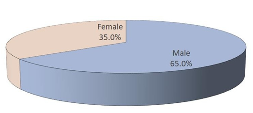
Out of a total of 60 cases, there were 39 men (65%) and 21 women (35%) in this study. Decreased hearing sensitivity and foul smelling otorrhea (86.7% each) were the most common presenting complaints followed by otalgia (50%), other complains are summarized in [Table 2]. On otoscopy findings, pars tensa abnormalities were seen in 55.8% while pars flacida abnormalities were seen in 61.7%. Among pars tensa abnormalities, 15% cases were having central perforation, followed by 8.3% cases having subtotal perforation. Other abnormalities include Total perforation, Grade 3 and 4 Retraction, Marginal perforation, Cholesteatoma etc.
Among pars flaccida abnormalities, cholesteatoma sac alone, cholesteatoma sac with grade 2 retraction, cholesteatoma sac with granulation tissue were seen. 53 patients had oedematous middle ear and 4 had polypoidal growth [Table 3]. Ossicular erosion was the most prevalent radiological finding in these scans (55%) and was followed by soft tissue mass (54.2%) and then scutum erosion (40.8%), as indicated in [Table 5]. Among minor findings were mastoid cortex dehiscence (5.8%), fallopian canal dehiscence (5%) and lateral semicircular canal fistula (1.7%).
Table 1
Age distribution of cases (n=60)
Table 2
Presenting complaints / Clinical findings
Table 3
Otoscopic Findings (n=120 ears)
Table 4
HRCT Findings (n=120 ears)
Table 5
Correlation of ossicular erosion with different clinicodemographic factors
Discussion
Recurring Otitis Media Cholesteatoma, an epithelial and fibrous stromal cystic lesion surrounded by an inflammatory response, is what delineates the squamosal form of cholesteatoma. 8 The middle ear is where temporal bone cholesteatoma typically develops, and it can result in catastrophic intracranial intrapetrous complications. 9
This severe condition is brought about by the keratinizing squamous epithelium that migrates from the external ear to the middle ear. 10 The most defining aspect of chronic otitis media is bone resorption.10 Imaging may be used to confirm the diagnosis in unusual presentations and is crucial for identifying cholesteatomas that has formed behind closed tympanic membrane.
Cholesteatomas are often diagnosed using otoscopy rather than imaging. By comparing the imaging results, the utility of HRCT to detect underlying diseases has typically been proven. Patients' ages ranged widely, from 13 to 60 years old, with a mean age of 26.03 years. The average age of patients’ having the disease was under 30 years for 75% of the patients'.
These results are consistent with observations made in several Indian studies, which showed that young people make up the bulk of cholesteatoma patients who require surgical intervention. The majority of them claimed that the largest occurrence was noted in people under 20 years of age. Similar findings were observed in a cross-sectional study conducted by Shami et al. on 50 patients of unsafe CSOM, was found to be male predominant disease with a mean age of 25.5 years.
In the current study, 65% of the patients were males. The ratio of men to women (M:F) was skewed. According to epidemiological research and earlier studies, the gender ratio is tilted towards male gender (M:F=1.46).
However, in different studies reviewed by us, the gender spectrum shows a variability. Kumari et al. in their study reported it to be 1.63, which is close to what is seen in the current study. 11 However, Sreedhar et al. in their study reported a much higher gender ratio of 2.71. 12 Decreased hearing sensitivity, foul smelling otorrhea and otalgia were the major presenting complaints affecting half or more of the patients. Blood-stained discharge (36.7%) was another major presenting complaint while complaints like tinnitus, dizziness and facial weakness were less common affecting 1.7 to 15% of the patients.
Traditionally, in CSOM, presenting complaints include persistent ear discharge and hearing loss. These in turn are responsible for subsequent complications of chronic otitis media.
Tak and Khilnani too in their study reported otorrhoea (80%) and hearing loss (66%) to be the most common presenting complaints and reported other complaints such as earache, headache, tinnitus, giddiness and facial paralysis in relatively much smaller proportion of patients affecting 4 to 18% of patients. 13
In the present study, on otoscopy, tympanic membrane abnormalities in 76 were seen in (62.5%) ears. Pars tensa and pars flaccida abnormalities were seen in 67 (55.8%) and 75 (61.7%) ears respectively. Middle ear could not be visualized in 119 subjects (99.2%). Findings suggestive of cholesteatoma formation were seen in 31 (25.8%) subjects.
Thukral et al too in their study reported otoscopic findings suggestive of in 54% of cases. 14 They also reported chronic otitis media– squamosal type in inactive phase CSOM in 20% cases and bilateral involvement in 24% patients. 14 Most of the other studies that have carried out HRCT evaluation of temporal bone abnormalities primarily include clinically or otoscopically suspected cases of cholesteatoma only. and did not describe the otoscopic findings in detail. Shaik et al. too in a recent study reported high prevalence of abnormal findings on otoscopic evaluation. They reported ear discharge (85.8%), scutum erosion (79.4%), attic retraction (62.8%) and cholesteatoma (32%) as the major findings. 15 As such, scutum erosion is difficult to be visualized on otoscopy and HRCT remains to be the confirmatory diagnosis for this purpose. The study of most common HRCT finding varies significantly in different studies.
Shah et al. (2022) in their study reported 100 patients with scutum erosion (64.28%), Koerner’s septum erosion (64.28%), eroded tegmen (35.71%), erosion of complete ossicular chain (57.14%) as the major HRCT findings helpful in guiding the surgical approach and treatment plan pre-operatively. 16 Ossicular erosion has also been reported as the major finding by Kumaresan and Nirmala in 87% cases who reported it to be the most common finding. 17
Both ossicular erosion as well as scutum erosion have been reported as the most common HRCT findings by Singh et al. in 90% and 84% cases. 18 In fact, in other contemporary studies too, ossicular erosion and/or scutum erosion has been reported as the most commonly detected HRCT abnormality in cases of cholesteatoma.
In the current study, most of the demographic factors and presenting complaints did not show a significant association with the HRCT findings. One of the reasons for this could be the fact that demographic profile and presenting complaints of CSOM patients are quite generalized and unspecific to the final diagnosis.
Moreover, progressive nature of these abnormalities shows a time dependence rather than a differential demographic profile of the patients. In the current study, a significant association of different types of HRCT abnormalities with some of the otoscopic findings was found. In the present study, absence of intact tympanic membrane was a feature that was significantly associated with presence of ossicular erosion, soft tissue mass, scutum erosion on HRCT.
In studies done before, an association of otoscopic findings with HRCT findings has also been reported which have shown the sensitivity and specificity for different HRCT abnormalities from 94.7% to 100% and from 50% to 100% respectively. 19
As such, despite these promising report in literature, it has been seen that HRCT offers a better opportunity to assess both the membranous as well as soft tissue changes vividly and clinical evaluation cannot undermine the role of HRCT. 20 Furthermore, studies to explore these relationships are recommended. HRCT is useful in diagnosing the middle ear pathologies effectively, particularly in assessment of damage caused to ossicular chain. 21 The major advantage of HRCT temporal bone is that it minimizes the role of invasive diagnostic and therapeutic modalities. 22 Treatment and management strategy formulation for middle ear pathologies can be successfully done with the help of HRCT findings. 23 HRCT is found to be cost-effective as compared to other invasive diagnostic modalities too. 24
Conclusion
Age of COM patients ranged from 13 to 60 years. Mean age was 26.03+-9.52 years and majority of patients ranged between 2nd to 3rd decade. Reduced hearing sensitivity and foul-smelling otorrhea were the most common presenting complaints. Among tympanic membrane abnormalities, Pars tensa abnormalities were seen in 55.8% and Pars flaccida abnormalities in 61.7% patients. On HRCT temporal bone ossicular erosion, was the most common finding 55% followed by soft tissue mass 54.2% and scutum erosion 40.8% as major finding. Minor HRCT finding included mastoid cortex dehiscense in 5.8% cases followed by fallopian canal dehiscence 5%. Significant association of Ossicular chain erosion, soft tissue mass and scutum erosion was seen with tympanic membrane abnormalities.
The findings of present study highlighted the fact that HRCT helps to diagnose the temporal bone abnormalities accurately which otherwise could not be differentiated clinically. Thus, findings of HRCT help in treatment planning and determining the need for surgical intervention.


