Indian Journal of Clinical Anatomy and Physiology
Indian Journal of Clinical Anatomy and Physiology (IJCAP) is an open access, peer-reviewed medical quarterly journal, published since 2014 under the auspices of the Innovative Education and Scientific Research Foundation (IESRF), which aims to uplift researchers, scholars, academicians, and professionals in all academic and scientific disciplines. we will be publishing articles ‘Ahead of Print’ immediately upon acceptance of manuscript. In addition, the journal allows free access (Open Access) to its contents, which is likely to attract more readers and citations to articles publishe...

A comparative study of cardio autonomic modulation in last term pregnancy with non-pregnant females
Abstract
Background: Pregnancy is associated with significant physiological adaptations in the cardiovascular system to maintain stable blood pressure despite increased blood volume, reduced blood viscosity, and the presence of the uteroplacental arteriovenous circulation. Aortocaval compression and enhanced sympathetic vasomotor activity, particularly during the third trimester, are considered major contributors to these hemodynamic changes. Increasing evidence suggests a strong asso iation between autonomic nervus system function and cardiovascular outcomes. Assessin cardiovascular autonomic modulation during late pregnancy may provide valuable insights into maternal cardiovascular adaptation and improve perinatal outcomes.
Materials and Methods: This prospective comparative study was conducted among pregnant women in the third trimester and age-matched non-pregnant women (18–30 years). Cardiovascular autonomic function was assessed using the CANWIN-504 system. The study compared autonomic modulation between the two groups to evaluate the physiological changes associated with late pregnancy.
Results: The study demonstrated alterations in cardiovascular autonomic modulation among women in the third trimester compared with non-pregnant women, indicating increased sympathetic activity and adaptive changes in autonomic regulation during late pregnancy. These findings suggest significant cardiovascular autonomic adaptations that support maternal and fetal physiological demands.
Conclusion: Third-trimester pregnancy is associated with significant changes in cardiovascular autonomic modulation, characterized by enhanced sympathetic activity as part of normal physiological adaptation. Assessment of autonomic function may serve as a useful tool for understanding maternal cardiovascular adjustments during pregnancy and may contribute to improved monitoring and better maternal and perinatal outcomes.
1. Introduction
The cardiovascular system's adaptation to pregnancy is significantly influenced by the autonomic nervous system (ANS).[1] At rest and in response to cardiovascular reflex stimulation, normal pregnancy is linked to an increase in sympathetic activity and a decrease in parasympathetic activity. It returns to baseline following delivery. The ideal uteroplacental blood flow is maintained by these modifications.[2][3]
The autonomic nervous system's function is crucial for both nourishing the developing fetus and circulatory adjustments throughout pregnancy.The sympathetic and parasympathetic nervous systems are the two separate anatomical and functional divisions of the autonomic nervous system.Systemic vascular resistance and blood pressure drop throughout the first half of pregnancy, but the resistance rises during the latter trimester.[4] A well controlled interaction between sympathetic and parasympathetic system is necessary for adapting the cardiovascular system to various hemodynamic needs not only under pathophysiological conditions like haemorrhage and shock, but also in physiological states such as pregnancy.[5]
While peripheral resistance and resting blood pressure drop during pregnancy, blood volue, heart rate, and cardiac output all rise.[6] Because the physiological hypervolemia of pregnancy would be expected to moderate substantial variations in blood pressure caused by a given stimulus, it may be assumed that baroreflex activity would be reduced during pregnancy.[7] Aortocaval compression brought on by an expanding gravid uterus significantly impairs venous return and cardiac output as gestational age rises, which causes a change in sympathetic nervous system activity toward an even stronger sympathetic and lower vagal modulation in the third trimester of pregnancy. During the third trimester of pregnancy, women with normal pregnancies have higher vasomotor sympathetic activity. In the last months of a typical pregnancy, there was a noticeable rise in sympathetic nerve activity, which assisted in bringing the arterial pressure back to normal levels. However, when this increase was too great, hypertension resulted. According to preliminary research findings, normal pregnancy may also be linked to an increase in resting vasomotor sympathetic outflow, and pregnancy itself may cause sympathetic activation even in the presence of normal blood pressure.[8]
The primary hypothesized cause of this alteration is aortocaval compression.Observations that shifting to an upright position could reduce sympathetic activation and that the delivery of a fetus resulted in the resumption of normal cardiovascular autonomic nerve activity within three months provided support for this notion.Changes in the autonomic nervous system that result in modifications to cardiac autonomic regulation are linked to all of these adaptations.Failure to make these adjustments could lead to pregnancyrelated problems such preeclampsia, eclampsia, and pregnancy-induced hypertension.
The cardiovascular reflex tests, including the orthostatic test, the isometric handgrip test, and heart rate variability measurement, allow for the non-invasive examination of autonomic nervous control. All the tests similarly disrupt the circulatory system.9 Analyzing the cardiovascular system's recovery can provide insights into the autonomic nervous regulation of hemodynamic.10 This study was helpful to investigate the changes in autonomic nervous system activity and its control over hemodynamic in last trimester pregnant females as compared to non-pregnant females.
2. Aims and Objective
The study was conducted to study about cardio autonomic imbalance amongst women of last trimester pregnancy and non pregnant women.
3. Materials and Methods
Non-invasive examination of autonomic nervous control can be performed using cardiovascular reflex assessments, including the Valsalva manoeuvre, the orthostatic test, the isometric test, and by assessing heart rate variability.[11]
3.1. Study design and settings
A comparative study was carried out amongst last trimester pregnant and non-pregnant women at Department of Physiology (Central Research Laboratory) of B.P.S. GMC (W), Sonepat, Haryana.
3.2. Study population
Sample size based upon proportion formula of two groups by taking prevalence in group 1: 90% and in group 2: 20% with estimated risk difference 7% at 95% compedence interval the required sample size is 86 subjects including 43 in each group.
3.3. Study conduct
All subjects were explained the procedure to be undertaken and a written consent will be obtained. All 43 pregnant women attending antenatal clinic were assessed for variation in sympathetic and parasympathetic system during last trimester of pregnancy (28-38 weeks) along with non-pregnant control group.
3.4. Inclusion criteria
18-30 years age limit. Healthy pregnant and non pregnant females. No history of any previous complicated pregnancy. No history of abortion.
3.5. Exclusion criteria
Twin gestation History of preeclampsia Gestational diabetes
3.6. Intervention
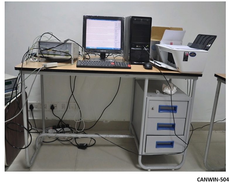
Cardiac autonomic function test for assessing cardio autonomic modulation (sympathetic and parasympathetic activity) was done by CANWIN-504(Cardiac Autonomic Neuropathy Analyser-504). CANWIN is a cutting-edge window-based computer equipped with a cardiac autonomic neuropathy (CAN) analysis system that includes interpretation. It maintains a comprehensive database to monitor subjects' histories for archival test access and comparisons. The Cardiac Autonomic Neuropathy Analyser model CANS 504 is a significant instrument for assessing and diagnosing autonomic dysfunctions by utilizing the R-R interval from electrograms and automated blood pressure readings.
3.7. Test for assessing parasympathetic activity include-
Resting heart rate: was calculated from ECG by using standard limb leads. Heart rate response to standing (30:15): was determined as the ratio of the R-R interval at beats 30 and 15 of the ECG taken immediately upon standing. This assessment measures the cardiovascular reaction triggered by transitioning from a horizontal to a vertical stance. The usual heart rate reaction to standing is significantly reduced by parasympathetic inhibition. Heart rate response to deep breathing: Heart rate was initially recorded during resting normal breathing and subsequently during deep breathing (6/min). The 3rd and 6th respiration of the ECG, along with the minimum R-R interval and the associated heart rate were determined.
Heart rate response to deep breathing: Heart rate was initially recorded during resting normal breathing and subsequently during deep breathing (6/min). The 3rd and 6th respiration of the ECG, along with the minimum R-R interval and the associated heart rate were determined.
3.8. Test for assessing Sympathetic activity-
Isometric hand grip exercise test: Before the exercise, subjects were allowed to rest for 20 minutes in a quiet room. Resting blood pressure of all subjects was measured by auscultatory method with help of mercury sphygmomanometer. First kortkoff sound indicated systolic blood pressure (SBP) and fifth kortkoff sound indicated diastolic blood pressure (DBP). Isometric handgrip test was done in both case and control group. After recording basal blood pressure, subjects were asked to perform isometric handgrip exercise. Subjects were told to hold the handgrip spring dynamometer in dominant hand to have full grip. Handles of dynamometer were compressed by subjects with maximum effort for few seconds. Then subject were told to perform 30% of maximum handgrip for 1 minute . During the test blood pressure was recorded from non exercising arm and again recorded after 5 minutes of exercise.[12]
3.9. Data collection and analysis plan
Statistical analysis: The collected data was entered in Microsoft Excel spread sheet. Mean +5d will be calculated for quantitative data. Percentage and proportion calculated for categorical data. Student’s’t’ test was used to find out the mean difference between the groups. Chi-square test was used to find out the association between the categorical data using SPSS (Version 220 software). AP value <0.005 were considered as statistically significant.
3.10. Ethical justification of study
The Study was conducted only after approval from Institutional Ethical Committee.
Confidentiality of all the patients is maintained in this study.
3.11. Expected outcomes
Autonomic imbalance in last trimester can leads to pregnancy induced hypertension which is a major obstetrical complication that results in adverse perinatal outcomes. Thus, timely identification of an autonomic imbalance during pregnancy can lessen the seriousness of complications and improves clinical results.
3.12. Future plans based on expected outcomes
CANS (Cardiac Autonomic Neuropathy System) will be an effective and automatic tool to assess autonomic cardiac dysfunction to prevent cardiac deaths, one of the most common cause of death in third trimester of pregnancy. Early diagnosis can save lives.
4. Results
The collected data was entered in Excel spreadsheet. Mean±SD (Standard Deviation) calculated for quantitative data, percentage. The Student’s ‘t’-test was applied to normally distributed variables to determine the mean difference utilizing Statistical Package for the Social Sciences software. P-value <0.005 is regarded as statistically significant.
|
Sympathetic test |
Control (43) Mean±SD |
Case (43) Mean±SD |
|---|---|---|
|
Sustained Hand grip (Change in diastolic BP) |
6.08 ± 6.27702 |
5.51 ± 11.08976 |
Table1 The mean score of Sustained hand grip in case group (5.51±11.08976) is lower than control group (6.08±6.27702), but the difference is considered statistically insignificant.
|
Parasympathetic test |
Control (43) Mean±SD |
Case (43) Mean±SD |
|---|---|---|
|
Resting Heart Rate(bpm) |
75.37 ± 4.89103 |
84.8 ± 15.72433 |
|
Deep breathing |
62.88 ± 22.05594 |
68.37 ± 36.56892 |
|
Standing (30:15 ratio) |
1.02 ± .31701 |
1.15 ± 1.43605 |
[Table 2] shows that 1. Mean score of Resting heart rate in case group (84.8±15.72433) is higher than control group (75.37±4.89103); and the difference is found to be statistically significant. 2. Means score of Deep breathing in case group (68.37±36.56892) is higher than control group (62.88±22.05594); but the difference is found to be statistically insignificant. 3. Mean score of Standing Blood pressure is higher in case group (1.15±1.43605) than control group (1.02±.31701); but the difference is found to be statistically insignificant.
|
Sympathetic test |
Control (43) Mean±SD |
Case (43) Mean±SD |
t-value |
p-value |
|---|---|---|---|---|
|
Sustained hand grip (change in diastolic BP) |
6.08±6.27702 |
5.51±11.08976 |
-0.304 |
0.762 |
|
Parasympathetic test |
Control (43) Mean±SD |
Case (43) Mean±SD |
t-value |
p-value |
|---|---|---|---|---|
|
Resting Heart Rate(bpm) |
75.37±4.89103 |
84.8±15.72433 |
3.838 |
.0001 |
|
Deep breathing |
62.88±22.05594 |
68.37±36.56892 |
.862 |
.391 |
|
Standing (30:15 ratio) |
1.02±.31701 |
1.15±1.43605 |
.607 |
.545 |
[Table 4]: shows that- 1. The mean difference in Resting heart rate among control and case group is statistically significant (P<0.005) 2. The mean difference in Deep breathing among control and case group is statically insignificant (P>0.005). 3. The mean difference in Standing Blood pressure among control and case group is statically insignificant (P>0.005).
5. Discussion
In the present study one test for accessing parasympathetic activity i.e. resting heart rate was found statistically significant in which the p-value calculated for mean result is <0.005. On comparison of resting heart rate of controls and cases it was found that the resting heart rate in last trimester pregnant women was higher as compared to non-pregnant females.Heiskanen Nonna et al suggest that parasympathetic deactivation may lead to higher heart rate and cardiac output during pregnancy.[2] So, the increase in heart rate in our study is consistent with reports of others.
The resting heart rate suddenly rises during the initial trimester of pregnancy, followed by a moderate increase later till term. However, in present study heart rate was measured only in 3rd trimester and it was higher in pregnant women when compared to control group.[13] Carson et al. also found a higher prevalence of tachycardia (29%) in healthy, asymptomatic patients in the third trimester of pregnancy. This result is consistent with the study of Heiskanen N, Sayers BM.[2][9] Kuo et al has reported thatspectral HRV analysis reveals a reduction in cardiac vagal activity and an increase in cardiac sympathetic activity during late pregnancy.[4]
With advancing gestational age, aorta caval compression from the growing pregnant uterus further impairs venous return and cardiac output, resulting in a change in autonomic nervous activity towards increased sympathetic and decreased vagal modulation in the third trimester of pregnancy.[12] During pregnancy, the mean maternal heart rate usually increases by an average of 10 to 20 beats per minute. The maternal heart rate begins to increases during the first few weeks of pregnancy and peaks in the late second to early third trimester.[15] After 32 weeks; the maintenance of cardiac output becomes more dependent on heart rate due to the fall in stroke volume.
The other tests performed on test and control group using the CANWIN-504, including test for accessing sympathetic activity ( Isometric Sustained hand grip exercise) and tests for accessing parasympathetic activity ( Standing Blood pressure and deep breathing exercise), the mean result obtained and the p-value obtained were not found to be statistically significant i.e. the p-value is >0.005. So, these tests are considered statistically insignificant.
Diastolic BP following isometric handgrip strength didn’t show any statistically significant change in last trimester pregnant women when compared with control group. These results were consistent with the study done by Barron et al.[16] In the late pregnancy, the mechanism of pressor response to isometric handgrip test is similar to post partum: there is an increase in cardiac output and reduction in forearm vascular resistance.[17]
The heart rate response to standing 30:15 was statistically non significant in last trimester pregnant and non pregnant women. These results coincide with the study done by Easterling et al. Doppler sonography, on the other hand has found that the decrease in both cardiac output and stroke volume caused by standing up are unaffected by pregnancy.[18] The vasoconstriction induced by orthostatic stress is diminished in pregnancy.[19] The forearm vasoconstrictor response is abolished during the last trimester of pregnancy compared with post partum.[17]
There was no notable difference in heart rate due to deep breathing between the case group and the control group. The current study aligns with the results of K E Juhani Airaksinen et al., as longitudinal studies initiated prior to pregnancy indicate that the maximal heart rate response to deep breathing steadily decreases during pregnancy.[20]
6. Conclusion
To conclude, In the present study, we conducted various sympathetic and parasympathetic tests to study the changes occurring in the last trimester of pregnancy (Between 18 to 30 years of age) due to cardio autonomic modulations. All tests carried out and analyzed on pregnant and non-pregnant females indicated that the differences in resting heart rate between the two groups were statistically significant. This research confirms that there is a physiological rise in resting heart rate during the final trimester of pregnancy compared to non-pregnant women.And CANWIN-504 is a potential tool to detect these changes in the autonomic nervous system and hence it can be beneficial to early diagnose the pathological state originating due to cardio autonomic imbalance in pregnant females and help prevent many serious pregnancy related cardio autonomic complications like pregnancy induced hypertension.
7. Summary
Cardiovascular modifications are necessary in pregnancy to maintain stable BP and to meet the oxygen and nutrients demands of both maternal and fetal organs. In our study it is found that there is significant increase in the resting heart rate towards the last trimester of pregnancy to ensure enough cardiac output to meet the needs of both expected mother and fetus.This cardiovascular-autonomic misbalance in the final trimester can result in pregnancy-induced hypertension, a significant obstetric complication that can cause serious maternal and fetal morbidity. Thus, the early identification of autonomic imbalance during pregnancy with CANWIN-504 can help lessen the severity of complications and enhance pregnancy clinical outcomes. So, the present study supported that CANS (Cardiac Autonomic Neuropathy System) will be an effective and automatic tool for early diagnosis of cardiac autonomic dysfunction to prevent pregnancy induced cardiac complications in last term pregnancy. Early diagnosis can save lives.
8. Source of Funding
None.
9. Conflict of Interest
None.
References
- Karumanchi S, Granger J. Preeclampsia and pregnancy related hypertensive disorders. Hypertension. 2016;67(2):238-42. [Google Scholar] [Crossref]
- Heiskanen N, Saarelainen H, Valtonen P, Lyyra-Laitinen T, Laitinen T, Vanninen E. Blood pressure and heart rate variability analysis of orthostatic challenge in normal human pregnancies. Clin Physiol Func Imag. 2008;28(6):384-90. [Google Scholar] [Crossref]
- Ekholm E, Piha S, Erkkola R, Antila K. Autonomic cardiovascular reflexes in pregnancy. A longitudinal study. Clin Autonomic Res. 1994;4(4):161-65. [Google Scholar] [Crossref]
- EKM, Piha S, Antila K, Erkkola R. Cardiovascular autonomic reflexes in mid pregnancy. Br J Obstetrics Gynaecology. 1993;100:177-182. [Google Scholar]
- Hans S. Autonomic function in normal pregnancy, the role of studying heart rate variability. Clin Sci. 2000;98(3):241-2. [Google Scholar] [Crossref]
- Hytten F, Leitch ITPohp. . Oxford. 1971. [Google Scholar]
- Thompson C, Tatro D, Convertino V. Baroreflex responses to acute changes in blood volume in humans. Am J Physiology. 1990;259:R792-8. [Google Scholar] [Crossref]
- Greenwood J, Scott E, Stoker J, Walker J, Mary D. Sympathetic neural mechanisms in normal and hypertensive pregnancy in humans. Circulation. 2001;104(18):2200-4. [Google Scholar] [Crossref]
- Sayers B. Analysis of heart rate variability. Ergonomics. 1972;16(1):17-32. [Google Scholar] [Crossref]
- Akselrod S, Gordon D, Madwed J, Snidman N, Shannon D, Cohen R. Hemodynamic regulation: Investigation by spectral analysis. Am J Physiol. 1985;249(4):H867-75. [Google Scholar] [Crossref]
- Wieling W. Non-invasive continuous recording of heart rate and blood pressure in the evaluation of Neurocardiovascular control. Autonomic failure. 1992. [Google Scholar]
- Kaur J, Batra A. Study of sympathetic andparasympathetic control of haemodynamics in mid pregnancy bycardiac autonomic neuropathy analyzer. Indian J Clin Anat Physiol. 2021;8(3):241-5. [Google Scholar] [Crossref]
- Ekholm E, Piha S, Antilla K, Erkkola R. Cardiovascular autonom ic reflexes in m idpregnanc y. Br J Obs tet Gynaecol. 1993;93(100):177-82. [Google Scholar] [Crossref]
- Kuo C. Chen G.Y., Yang M.J., Tsai Y.S.: "The effect of position on autonomic nervous activity in late pregnancy". Anaesthesia. 1997;52(12):1161-1165. [Google Scholar] [Crossref]
- Easterling T, Benedetti T, Schmucker B, Millard S. Maternal hemodynamics in normal and preeclamptic pregnancies: a longitudinal study. Obstet Gynecol. 1990;76:1061-9. [Google Scholar]
- Barron W, Mujais S, Zinaman M, Bravo E, Lindheimer M. Plasma catecholamine responses to physiologic stimuli in normal human pregnancy. Am J Obstet Gynecol. 1986;154(1):80-4. [Google Scholar] [Crossref]
- Nisell H, Hjemdahl P, Linde B, Lunell N. Sympathoadrenal and cardiovascular reactivity in pregnancy induced hypertension. I. Responses to isometric exercise and a cold pressor test. BJOG:. An Int J Obstet Gynaecol. 1985;92(7):722-31. [Google Scholar] [Crossref]
- Easterling T, Schmucker B, Benedetti T. The hemodynamic effects of orthostatic stress during pregnancy. Obstet Gynecol. 1988;72(4):550-2. [Google Scholar]
- Clark S, Cotton D, Pivarnik J, Lee W, Hankins G, Benedetti T, et al. Position change and central hemodynamic profile during normal third-trimester pregnancy and post partum. Am J Obstet Gynecol. 1991;164(3):883-7. [Google Scholar] [Crossref]
- Airaksinen K, Salmela PI IM, Kirkinen P, Linnaluoto M, Takkunen J. Effect of pregnancy on autonomic nervous function and heart rate in diabetic and nondiabetic women. Diab Care. 1987;10(6):748-51. [Google Scholar] [Crossref]
- Abstract
- 1. Introduction
- 2. Aims and Objective
- 3. Materials and Methods
- 3.1. Study design and settings
- 3.2. Study population
- 3.3. Study conduct
- 3.4. Inclusion criteria
- 3.5. Exclusion criteria
- 3.6. Intervention
- 3.7. Test for assessing parasympathetic activity include-
- 3.8. Test for assessing Sympathetic activity-
- 3.9. Data collection and analysis plan
- 3.10. Ethical justification of study
- 3.11. Expected outcomes
- 3.12. Future plans based on expected outcomes
- 4. Results
- 5. Discussion
- 6. Conclusion
- 7. Summary
- 8. Source of Funding
- 9. Conflict of Interest
- References
Article Metrics
- Visibility 201 Views
- Downloads 129 Views
- DOI 10.18231/j.ijcap.16832.1779172535
-
CrossMark

- Citation
- Received Date April 11, 2026
- Accepted Date May 13, 2026
- Publication Date June 30, 2026