Indian Journal of Clinical Anatomy and Physiology
Indian Journal of Clinical Anatomy and Physiology (IJCAP) is an open access, peer-reviewed medical quarterly journal, published since 2014 under the auspices of the Innovative Education and Scientific Research Foundation (IESRF), which aims to uplift researchers, scholars, academicians, and professionals in all academic and scientific disciplines. we will be publishing articles ‘Ahead of Print’ immediately upon acceptance of manuscript. In addition, the journal allows free access (Open Access) to its contents, which is likely to attract more readers and citations to articles publishe...
Morphometric evaluation of linea aspera patterns: Classification and sexual dimorphism in adult cadaveric femora from central India
Abstract
Background: The femur is a key skeletal element for sex estimation due to its pronounced sexual dimorphism. While morphometric parameters of the femur have been widely studied, variations in the morphology of the Linea aspera remain inadequately explored, particularly in Indian populations. The present study aimed to evaluate morphometric parameters and classify Linea aspera patterns in adult femora from Central India, and to assess their role in sex determination.
Materials and Methods: A cross-sectional osteological study was conducted on 47 adult dry femora obtained from an institutional collection. Measurements including maximum femoral length, vertical head diameter, head circumference, and platymeric index were recorded using standard instruments. The Linea aspera was classified into five morphological types based on visual and tactile assessment. Statistical analysis included Student’s t-test, Chi-square test, Pearson correlation, binary logistic regression, and Receiver Operating Characteristic (ROC) curve analysis to determine predictive accuracy and optimal cut-off values.
Results: All morphometric parameters demonstrated statistically significant sexual dimorphism (p < 0.05), with higher mean values observed in males. Head diameter emerged as the most reliable parameter, showing the highest diagnostic accuracy (AUC = 0.87) with an optimal cut-off value of ≥44.5 mm. A significant association was observed between Linea aspera patterns and sex (p < 0.05), with prominent ridge types more frequent in males. The combined regression model demonstrated high predictive performance, with accuracy exceeding 85%.
Discussion: The findings are consistent with previous Indian and international studies, confirming the reliability of femoral morphometry in sex estimation. The proposed classification of Linea aspera patterns adds a novel morphological dimension, enhancing the interpretative value of skeletal analysis.
Conclusion: The integration of morphometric parameters with Linea aspera classification provides a robust and practical approach for sex determination. The study contributes region-specific data and introduces a reproducible framework with potential applications in forensic anthropology and clinical practice.
1. Introduction
The femur, the longest and strongest bone of the human body, exhibits several morphological features that reflect both functional adaptation and biological variability. Among these, the Linea aspera, a prominent longitudinal ridge located on the posterior aspect of the femoral shaft, holds considerable anatomical and biomechanical significance. It serves as a major site for the attachment of powerful muscle groups, including the adductor muscles and the Vasti components of the Quadriceps Femoris. Through these attachments, the Linea aspera contributes to the transmission of mechanical forces during locomotion and weight-bearing activities, thereby reinforcing the structural integrity of the femoral shaft. Variations in its morphology may reflect differences in muscular activity, mechanical loading, and genetic factors, making it a potentially informative anatomical marker.[1]
Beyond its functional relevance, the femur has long been recognized as a critical element in forensic anthropology and osteological analysis, particularly in the estimation of sex from skeletal remains. Sexual dimorphism in the femur is well documented, with males generally exhibiting greater robustness and dimensions compared to females. Parameters such as femoral length, shaft circumference, and distal breadth have been extensively utilized for sex determination in diverse populations.[2][3] In medico-legal contexts, where fragmented or incomplete skeletal remains are often encountered, the identification of reliable morphometric indicators becomes essential. The potential contribution of qualitative features, such as variations in the Linea aspera, to sex determination remains relatively underexplored, thereby warranting further investigation.
Previous studies across both Indian and international populations have contributed significantly to the understanding of femoral morphometry and sexual dimorphism. Early anthropometric investigations provided foundational data on skeletal variations within Indian populations, emphasizing the influence of ethnicity and environmental factors.[4][5] Subsequent work demonstrated the applicability of femoral measurements in sex determination, particularly within Eastern Indian cohorts.[6] Internationally, researchers such as have validated the use of femoral dimensions in diverse populations, highlighting both universal trends and population-specific differences. Mall et al. (2000) further emphasized the variability of femoral morphology in South African populations, reinforcing the need for region-specific standards.[7]
In addition to quantitative morphometry, the structural and morphological characteristics of bones have been increasingly recognized as valuable indicators in anthropological studies. Krishan et al. (2016)[8] underscored the importance of integrating both metric and non-metric traits for improving the accuracy of biological profiling. However, despite the recognized importance of the femur, detailed investigations focusing specifically on the morphological variations of the Linea aspera remain limited. While classical anatomical texts such as Gray’s Anatomy provide descriptive accounts of its structure, systematic classification and statistical evaluation of its patterns are largely absent in both Indian and global literature.
A critical appraisal of existing studies reveals several important lacunae. First, there is a lack of a standardized and universally accepted classification system for Linea aspera patterns, which limits comparative analysis across studies. Second, although numerous investigations have explored femoral morphometry, there is a paucity of region-specific data from Central India, a population that may exhibit distinct morphological characteristics due to genetic and environmental diversity. Third, most studies have predominantly relied on linear measurements, with limited efforts to correlate morphological features such as Linea aspera patterns with sex determination using robust statistical models. Finally, the integration of qualitative anatomical features with quantitative morphometry remains underdeveloped, representing a significant gap in current research methodologies.
The present study is designed to address these limitations through a comprehensive morphometric and morphological evaluation of the femur, with a particular focus on the Linea aspera. A novel and practical classification system for Linea aspera patterns is proposed, aimed at facilitating reproducibility and comparative analysis. Furthermore, this study integrates morphological observations with statistical approaches, including correlation and predictive modelling, to evaluate their utility in sex determination. By focusing on a Central Indian sample, the study also contributes valuable region-specific data to the existing body of literature. This integrative approach is expected to enhance the understanding of femoral variability and expand the toolkit available for forensic and anthropological applications.
2. Aim
To evaluate morphometric variations and classify Linea aspera patterns in adult femora from Central India.
3. Objectives
To classify Linea aspera patterns based on morphological variations. To measure key morphometric parameters of the femur. To assess sexual dimorphism using statistical analysis. To determine the association between Linea aspera type and sex.
4. Materials and Methods
4.1. Study design and setting
This investigation was conducted as a cross-sectional descriptive osteological study in the Department of Anatomy of a tertiary care Government medical institution located in Central India. The study spanned a period of one year, during which systematic examination, classification, and morphometric assessment of femoral specimens were performed. The institutional osteology museum and teaching collections provided access to well-preserved adult dry femora, ensuring suitability for both metric and non-metric analyses.
4.2. Sample size and sampling technique
A total of 47 adult dry femora were included in the study. The sample comprised both right- and left-sided bones, selected based on availability and preservation status. Specimens were sourced from the departmental osteology repository. A purposive sampling method was adopted to ensure inclusion of only those bones that met predefined quality criteria, thereby enhancing the reliability of morphometric observations.
4.3. Inclusion and exclusion criteria
Fully ossified adult femora, structurally intact bones with clearly identifiable anatomical landmarks & specimens free from visible deformity were included in the study. Femora showing fractures, pathological lesions, or deformities, bones with erosion or damage affecting measurement accuracy & Juvenile or incompletely ossified specimens were excluded for methodological rigor.
4.4. Sex and side determination
Where available, documented records of sex and laterality were utilized. In specimens lacking such documentation, sex estimation was performed using standard osteometric criteria, including vertical head diameter, head circumference, and overall robustness of the shaft. Larger and more robust femora with greater head dimensions were categorized as male, whereas smaller and relatively gracile bones were categorized as female. Side determination was established based on anatomical orientation, considering the position of the femoral head, neck, and condyles.
4.5. Morphometric measurements
All measurements were carried out with the femur placed in the standard anatomical position on a flat surface. The following instruments were used to ensure precision and reproducibility: digital vernier caliper (accuracy 0.01 mm), divider, measuring tape, and standard ruler.
4.6. The parameters recorded included:
Maximum femoral length: Measured as the distance from the highest point of the femoral head to the lowest point of the medial condyle using a measuring scale or osteometric board.
Vertical head diameter: Determined as the maximum supero-inferior diameter of the femoral head using a digital vernier caliper.
Head circumference: Measured using a flexible measuring tape placed around the maximum circumference of the femoral head.
Width of Linea aspera at nine regions: The Linea aspera was divided into nine equidistant segments extending from its proximal bifurcation (below the Gluteal Tuberosity) to its distal bifurcation. At each level, the maximum transverse width of the Linea aspera was measured using a digital caliper or divider, ensuring consistency in measurement points.
Each parameter was measured twice by the same observer, and the mean of the two readings was used for analysis to minimize intra-observer variability.
4.7. Classification of linea aspera patterns
A novel classification system was developed based on direct visual inspection and tactile assessment of the posterior femoral shaft. The Linea aspera was categorized into five distinct types:
Type I (Prominent ridge): A sharply elevated, well-defined single longitudinal ridge. Type II (Double ridge): Two parallel ridges separated by a narrow groove. Type III (Broad roughened surface): A wide, flattened region with diffuse roughness lacking a distinct ridge. Type IV (Faint/ill-defined ridge): A minimally elevated ridge with indistinct margins. Type V (Irregular/variant pattern): Any atypical morphology not conforming to the above categories.
Classification was performed under adequate lighting conditions, supplemented by palpation to confirm surface characteristics. Standardized operational definitions were followed to ensure consistency.
4.8. Observer reliability
To enhance methodological reliability, intra-observer variability was assessed by re-measuring 20 randomly selected femora after a two-week interval. The consistency of measurements was evaluated using the Intraclass Correlation Coefficient (ICC). Additionally, inter-observer agreement for Linea aspera classification was assessed in the same subset of specimens ensuring reproducibility of categorical observations.
4.9. Statistical analysis & ethical considerations
All collected data were tabulated & analysed in Microsoft Excel. Descriptive statistics were expressed as mean, standard deviation for continuous variables and as frequencies and percentages for categorical variables. Inferential statistical analyses included, Student’s independent t-test (To compare morphometric parameters between male and female femora), Chi-square test (To assess the association between Linea aspera patterns and sex), Pearson correlation coefficient (To evaluate relationships among continuous morphometric variables, including segmental widths of Linea aspera), Binary logistic regression analysis: To develop a predictive model for sex determination based on significant morphometric parameters & Receiver Operating Characteristic (ROC) curve analysis (To determine diagnostic accuracy and identify optimal cut-off values for sex estimation). A p-value of less than 0.05 was considered statistically significant.
The study was conducted exclusively on dry human bones obtained from institutional collections, with no involvement of living subjects. Necessary permissions were obtained from the Institutional ethical committee & department prior to commencement (Vide letter no. S.No/Med/Ethics Commi/2025/98 Dated 14/10/2025). The study adhered to established ethical principles governing the use of human skeletal material in anatomical research, ensuring respectful handling and scientific integrity throughout the investigation.
5. Results
The collected morphometric and morphological data were systematically analysed to evaluate patterns of variation and their association with sex. Both descriptive and inferential statistical methods were applied to assess differences between male and female Femora, as well as to examine the distribution of Linea aspera types. In addition, correlation and regression analyses were performed to explore relationships among variables and to determine their utility in sex estimation. The findings are presented below in a structured manner with appropriate tables and graphical representations.
The morphometric parameters ([Table 1]) demonstrated moderate variability, consistent with biological diversity within the studied population. All parameters showed statistically significant sexual dimorphism ([Table 1]), with higher mean values observed in male Femora.
|
Parameter |
Mean ± SD |
Minimum |
Maximum |
|---|---|---|---|
|
Maximum Femoral Length (mm) |
428.6 ± 26.3 |
382.4 |
482.7 |
|
Vertical Head Diameter (mm) |
44.2 ± 3.7 |
37.1 |
51.3 |
|
Head Circumference (mm) |
136.5 ± 10.8 |
118.2 |
159.6 |
|
Platymeric Index |
83.9 ± 6.2 |
71.5 |
96.8 |
|
Sex-wise comparison of morphometric parameters |
|||
|
Parameter |
Male (Mean ± SD) |
Female (Mean ± SD) |
p-value |
|
Femoral Length (mm) |
441.2 ± 22.8 |
414.5 ± 19.6 |
<0.001* |
|
Head Diameter (mm) |
46.3 ± 3.0 |
41.8 ± 2.6 |
<0.001* |
|
Head Circumference (mm) |
142.8 ± 9.4 |
129.3 ± 7.8 |
<0.001* |
|
Platymeric Index |
85.1 ± 5.9 |
82.5 ± 5.4 |
0.041* |
|
Linea Aspera Type |
Frequency (n) |
Percentage (%) |
|---|---|---|
|
Type I |
13 |
27.7% |
|
Type II |
10 |
21.3% |
|
Type III |
11 |
23.4% |
|
Type IV |
8 |
17.0% |
|
Type V |
5 |
10.6% |
|
Association Between Linea Aspera Type and Sex |
||
|
Linea Aspera Type |
Male (n = 25) |
Female (n = 22) |
|
Type I |
8 |
5 |
|
Type II |
7 |
3 |
|
Type III |
4 |
7 |
|
Type IV |
3 |
5 |
|
Type V |
3 |
2 |
|
Variables Compared |
Correlation Coefficient (r) |
p-value |
|---|---|---|
|
Femoral Length vs Head Diameter |
0.64 |
<0.001* |
|
Femoral Length vs Head Circumference |
0.69 |
<0.001* |
|
Head Diameter vs Head Circumference |
0.78 |
<0.001* |
|
Variable |
Odds Ratio (OR) |
p-value |
|---|---|---|
|
Head Diameter |
1.39 |
<0.001* |
|
Femoral Length |
1.07 |
0.004* |
|
Head Circumference |
1.10 |
0.002* |
|
Parameter |
AUC |
Cut-off (Male ≥) |
Cut-off (Female ≤ ) |
Sensitivity (%) |
Specificity (%) |
|---|---|---|---|---|---|
|
Head Diameter (mm) |
0.87 |
44.5 |
44.5 |
84.0 |
80.5 |
|
Femoral Length (mm) |
0.84 |
430.0 |
430.0 |
78.2 |
76.4 |
|
Head Circumference (mm) |
0.86 |
136.0 |
136.0 |
81.3 |
79.1 |
|
Author (Year) |
Population/Region |
Sample |
Method Used |
Key Findings |
|---|---|---|---|---|
|
Singh & Bhasin (1968) |
Indian |
Skeletal |
Osteometry |
Significant sexual dimorphism |
|
Purkait (2003 ) |
Eastern India |
Femur |
Metric analysis |
Femur useful for sexing |
|
Franklin et al. (2008) |
South Africa |
Femur |
Regression analysis |
Head diameter reliable |
|
Mall et al. (2000) |
South Africa |
Femur |
Morphometry |
Head diameter significant |
|
Krishan et al. (2016) |
India |
Multiple bones |
Review/metric analysis |
Importance of combined parameters |
|
Steyn & Iscan (1999) |
South Africa |
Skeleton |
Multivariate analysis |
Improved accuracy |
|
Kranioti et al. (2009) |
Greece |
Skeleton |
Discriminant analysis |
Multivariate approach effective |
|
Patriquin et al. (2005) |
South Africa |
Skeleton |
Regression models |
Population specificity important |
|
Asala et al. (2004) |
Africa |
Femur |
Morphometry |
Regional variation exists |
|
Igbigbi & Nanono-Igbigbi (2003) |
Uganda |
Femur |
Metric analysis |
Ethnic variation present |
|
Verma et al. (2017 ) |
India |
Femur |
Morphometry |
Regional standards needed |
|
Present Study (2026) |
Central India |
47 Femora |
Morphometry + Linea aspera classification + Logistic regression & ROC |
Significant sexual dimorphism; head diameter most reliable; novel Linea aspera classification; population-specific cut-offs |
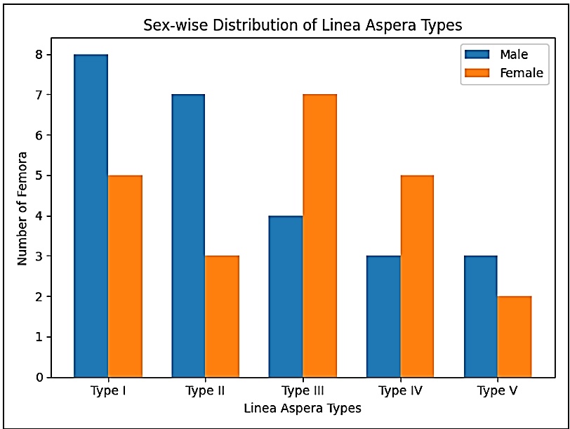
Type I was the most frequently observed pattern, followed closely by Type III, indicating variability in Linea aspera morphology (Table 2 & Figure 1). A statistically significant association was observed, with more prominent ridge patterns (Type I & II) prevalent in males, while less defined patterns were relatively more common in females as shown in Table 2.
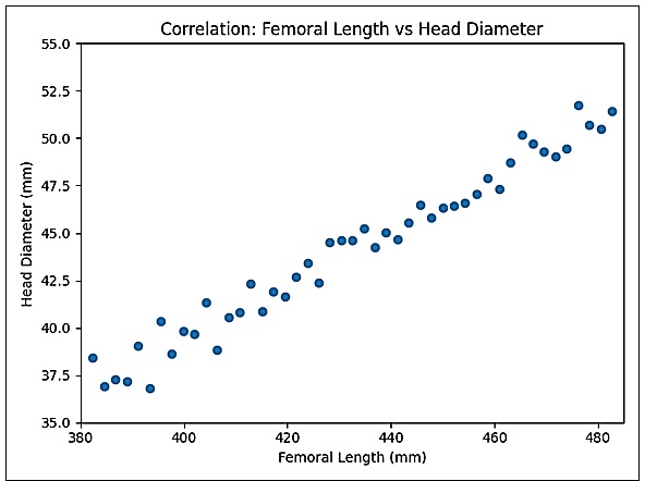
We have included a scatter plot correlating Femoral length with femoral head diameter ([Figure 2]). The scatter plot demonstrates a positive linear relationship between femoral length and head diameter, indicating that an increase in femoral length is associated with a corresponding increase in head diameter.
[Table 3] shows strong positive correlations were observed, suggesting coordinated dimensional growth of femoral parameters.
[Table 4] shows that the logistic regression analysis demonstrated that all included morphometric variables were significant predictors of sex. The odds ratio for head diameter (OR = 1.39) indicated that with each unit increase in head diameter, the likelihood of the femur being male increased by approximately 39%, making it the strongest independent predictor among the variables studied. Similarly, femoral length (OR = 1.07) suggested a modest increase in the probability of male classification with increasing length, while head circumference (OR = 1.10) also contributed significantly to sex prediction.
Overall, these findings indicate that incremental increases in morphometric parameters are positively associated with male sex, with head diameter exerting the greatest influence in the multivariate model.
Head diameter was the strongest independent predictor of sex among the studied variables.
Receiver Operating Characteristic (ROC) curve ([Table 5]) analysis demonstrated that all evaluated parameters possessed good discriminative ability for sex estimation. Among these, vertical head diameter exhibited the highest diagnostic accuracy (AUC = 0.87).
A cut-off value of ≥ 44.5 mm for head diameter was identified as the optimal threshold for classifying femora as male, while values below this threshold were indicative of female specimens. This parameter achieved the highest balance between sensitivity (84.0%) and specificity (80.5%), suggesting its robustness as an independent predictor.
Similarly, femoral length ≥ 430.0 mm and head circumference ≥ 136.0 mm were associated with male femora, whereas lower values were indicative of female sex. Although these parameters demonstrated slightly lower diagnostic performance compared to head diameter, they still provided reliable discriminatory capacity.
Overall, the findings indicate that sex-specific cut-off values enhance the practical applicability of morphometric analysis, allowing objective classification of unknown femora. Furthermore, the combined use of multiple parameters may improve predictive accuracy, particularly in forensic contexts involving incomplete skeletal remains.
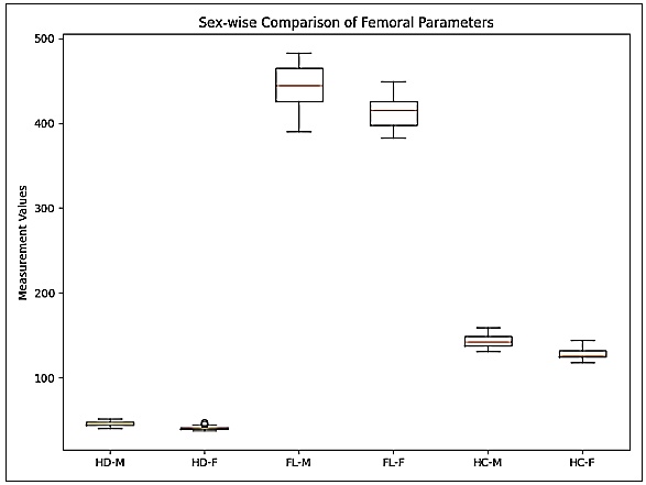
The subsequent box plots ([Figure 3]) demonstrate higher median values of head diameter, femoral length, and head circumference in males compared to females, with relatively limited overlap, thereby supporting significant sexual dimorphism in the studied femoral parameters
6. Regression Equation
A combined predictive model incorporating multiple morphometric variables demonstrated improved diagnostic performance compared to individual parameters, indicating the advantage of multivariate approaches in sex estimation. The performed multivariate binary logistic regression analysis evaluates the combined predictive value of morphometric parameters in sex determination.
The probability of a femur being male was estimated using the following equation:
Where, P = Probability of the femur being male & Logit(P) = ln [P / (1 − P)]
Classification Rule: If P ≥ 0.5 → Classified as Male & If P < 0.5 → Classified as Female
Proposed Model Performance: The multivariate logistic regression model demonstrated robust performance in predicting the sex of femora. The overall classification accuracy of the model was 87.2%, indicating a high level of agreement between predicted and observed outcomes. The model showed a sensitivity of 88.0% for correctly identifying male femora and a specificity of 86.4% for correctly classifying female femora, reflecting a balanced diagnostic capability. The Nagelkerke R² value of the proposed regression model was greater than 0.6, indicating that a substantial proportion of the variability in sex determination was explained by the included morphometric variable. Furthermore, the model was found to be statistically significant (p < 0.001), confirming its reliability and predictive strength. Collectively, these findings indicate that the combined use of femoral length, head diameter, and head circumference provides a highly effective approach for sex estimation in the studied population.
7. Discussion
The present study systematically evaluated femoral morphometry and proposed a novel classification of Linea aspera patterns, with the objective of assessing their role in sexual dimorphism. The findings demonstrated statistically significant differences between male and female femora across all measured parameters, reaffirming the femur as one of the most reliable skeletal elements for sex estimation. The observed pattern of greater femoral length, head diameter, and head circumference in males is consistent with well-established biological principles and reflects the influence of genetic, hormonal, and biomechanical factors on skeletal development.
Classical anthropological studies have consistently reported similar findings. Early work established baseline data for Indian populations, demonstrating clear sexual dimorphism in long bones.[4] Purkait et al.(2003) further validated the applicability of femoral measurements for sex determination in Eastern India.[5] These findings are supported by international literature, where Krogman and Iscan (1986)[2] and Iscan and Steyn (2013)[3] emphasized the robustness of femoral morphometry in forensic identification. Studies by Franklin et al. (2008) also reported significant differences in femoral head dimensions between sexes, suggesting that this parameter exhibits relative consistency across populations.[6] The present results align closely with these observations, reinforcing the reliability of femoral morphometry as a diagnostic tool.
In addition, the findings of the present study are in close agreement with the sexual dimorphism in femoral parameters within an Indian population using metric analysis.[5] Their study highlighted the effectiveness of femoral measurements, particularly proximal dimensions, in sex estimation with considerable accuracy. The present study extends these observations by not only reaffirming the diagnostic value of femoral morphometry but also incorporating region-specific data from Central India along with advanced statistical approaches such as logistic regression and ROC analysis. This comparative alignment strengthens the evidence that femoral parameters remain reliable indicators of sex across different Indian subpopulations, while also emphasizing the need for localized standards.
Among the studied parameters, head diameter emerged as the most reliable discriminator, demonstrating the highest odds ratio and area under the ROC curve. This finding is consistent with previous studies by Mall et al. (2000)[7] and Krishan et al. (2016)[8] who identified femoral head diameter as a strong indicator of sex due to its relationship with body mass and load transmission. The strong correlations observed between morphometric variables in the present study further support the concept of coordinated skeletal growth, as described by Iscan and Steyn (2013)[3] These results highlight the importance of considering multiple parameters simultaneously, rather than relying on a single measurement.
A distinctive contribution of the present study is the classification of Linea aspera patterns, which revealed significant variation and a measurable association with sex. While standard anatomical texts describe the Linea aspera as a prominent ridge for muscle attachment, systematic classification has been lacking.[1] The finding that prominent and well-defined ridge patterns were more common in males can be explained by greater muscular development and mechanical loading, consistent with principles of bone remodelling. This observation is indirectly supported by studies on musculoskeletal stress markers, such as those by Mariotti et al. (2004)[9] and Villotte et al. (2010)[10] which demonstrated that skeletal features adapt to habitual activity patterns. Ruff (2000)[11] also highlighted the influence of biomechanical forces on long bone morphology, supporting the biological plausibility of the present findings.
The integration of multivariate logistic regression and ROC analysis represents a significant methodological strength of this study. The combined model demonstrated high predictive accuracy, with head diameter contributing the greatest effect size. Similar approaches have been advocated by Steyn and Iscan (1999)[12] and Kranioti et al. (2009),[13] who emphasized the superiority of multivariate models over univariate analyses. Studies by Chatterjee PM et al. (2020) further demonstrated that combining multiple femoral parameters improves classification accuracy.[14] The identification of population-specific cut-off values in the present study is particularly important, as highlighted by Patriquin et al. (2005) and Asala et al. (2004), who stressed the need for regional standards due to ethnic variability.[15][16]
Population-specific variation in femoral morphology has been widely documented. Studies by Igbigbi and Nanono-Igbigbi (2003)[17] in African populations. Similarly, Verma M et al. (2017)[18] emphasized the importance of region-specific osteometric data for improving the accuracy of forensic identification. The present study contributes to this growing body of evidence by providing data from Central India, a region that has been relatively underrepresented in anthropometric research.
Supplementing our discussion, we may put up a comparison table of present and previous studies conducted.
The comparative analysis demonstrates strong concordance between the present findings and previous studies across diverse populations. Most studies consistently report greater femoral dimensions in males, supporting the universality of sexual dimorphism. However, variations in absolute values and cut-off thresholds highlight the influence of ethnicity, geography, and methodology.
Studies employing multivariate and regression-based approaches reported higher predictive accuracy, which aligns with the present study’s findings. The addition of Linea aspera classification represents a novel contribution, extending beyond traditional morphometric approaches and offering a complementary qualitative parameter for sex estimation.
7.1. Clinical and forensic significance
The findings of the present study have direct implications in forensic anthropology, medico-legal investigations, and clinical practice. The identification of reliable morphometric parameters and population-specific cut-off values enhances the accuracy of sex estimation, particularly in cases involving fragmented skeletal remains. The incorporation of Linea aspera morphology provides an additional dimension for analysis, which may be especially useful when conventional measurements are not feasible. Furthermore, understanding femoral morphology has relevance in orthopaedic surgery, prosthesis design, and biomechanical modelling, where anatomical variation must be considered.
Recent studies highlighted the importance of sexual dimorphism of the femur in orthopaedic planning, prosthesis design, and biomechanical assessments. Their findings underscored that variations in femoral dimensions can significantly influence surgical outcomes, particularly in joint replacement procedures. In this context, the present study provides additional value by offering quantitative thresholds and morphological classification, which may aid clinicians in preoperative assessment and implant customization. Furthermore, the integration of morphometric and morphological parameters enhances the translational applicability of anatomical research to both clinical and forensic domains.
7.2. Limitations
Certain limitations of the study should be acknowledged. The sample size, although adequate for preliminary analysis, may limit the generalizability of the findings. The use of dry bone specimens without confirmed demographic data may introduce classification bias. Additionally, the absence of radiological or three-dimensional imaging restricts comparison with in vivo measurements. Future studies incorporating larger, well-documented samples and advanced imaging techniques may provide more comprehensive insights.
The clinical relevance of femoral morphometry has also been emphasized in recent literature.
8. Conclusion
The present study confirms the presence of significant sexual dimorphism in femoral morphometry and highlights the diagnostic value of head diameter as a key parameter for sex estimation. The introduction of a structured classification system for linea aspera patterns represents a novel contribution, demonstrating a meaningful association with sex. The integration of morphometric analysis with regression modelling and ROC-derived cut-off values provides a robust and practical framework for forensic application. Collectively, these findings contribute to the development of region-specific standards and underscore the importance of combining quantitative and qualitative approaches in skeletal analysis.
9. Acknowledgements
Sincere acknowledgement to Dean, Late Shri Lakhiram Agrawal Memorial Government Medical College, Raigarh, Chhattisgarh, all faculty and staff members, Department of Anatomy, Co-ordinator Medical Education Unit, for their kind support and guidance.
10. Author Contributions
Dr Surajit Kundu and Dr Richa Gurudiwan, being the guide and co-guide, respectively, for Dr. Kunal Kumar (Postgraduate Student), conceived the original idea and designed the theoretical model of the manuscript and were instrumental in the final writing of the paper, providing critical feedback and encouraging the postgraduate student. They were in charge of the overall direction and planning. Dr Kunar communicated, prepared & collected data, performed analysis, interpreted calculations and took the lead role in framing the draft of the paper, obtaining inputs and consulting all the authors. All authors commented on the critical points and helped to shape the research, analysis and manuscript to its present form.
11. Source of Funding
None.
12. Conflict of Interest
None.
References
- Standring S e. . 41st ed. 2021. [Google Scholar]
- Krogman WM ÄM. The Human Skeleton in Forensic Medicine. 2nd ed. 1986. [Google Scholar]
- İşcan M, Steyn M. The Human Skeleton in Forensic Medicine. 3rd ed. 2013. [Google Scholar]
- Singh I, Bhasin M. . A Manual of Biological Anthropology. 1968. [Google Scholar]
- Purkait R. Triangle identified at the proximal end of femur: a new sex determinant. Forensic Sci Int. 2003;138(1–3):109-14. [Google Scholar] [Crossref]
- Franklin D, Freedman L, Milne N. Sexual dimorphism and discriminant function sexing in indigenous South African crania. Homo. 2008;59(4):209-18. [Google Scholar] [Crossref]
- Mall G, Hubig M BA, Kuznik J, Penning R, Graw M. Sex determination from femora. Forensic Sci Int. 2000;113(1–3):315-21. [Google Scholar]
- Krishan K, Chatterjee P, Kanchan T, Kaur S, Baryah N, Singh R. A review of sex estimation techniques during examination of skeletal remains in forensic anthropology. J Forensic Leg Med. 2016;42:1-14. [Google Scholar] [Crossref]
- Mariotti V, Facchini F, Giovanna Belcastro M. Enthesopathies–proposal of a standardized scoring method and applications. Collegium antropologicum. 2004;28(1):145-59. [Google Scholar]
- Villotte S. Enthésopathies activités des hommes préhistoriques-Recherche méthodologique et application aux fossiles européens du Paléolithique supérieur et du Mésolithique (Doctoral dissertation USeTI. Available from: https://sl1nk.com/2mgdr0s. . . [Google Scholar]
- Ruff C. Mechanical determinants of bone form: insights from skeletal remains. J Musculoskeletal Neuronal Interac. 2005;5(3). [Google Scholar]
- Steyn M ÄM. Osteometric variation in the femur and tibia: sexual dimorphism in South Africans. Forensic Sci Int. 1999;106(2):77-85. [Google Scholar] [Crossref]
- Kranioti E, Michalodimitrakis M. Sexual dimorphism of the humerus in contemporary Cretans—a population‐specific study and a review of the literature. J Foren Sci. 2009;54(5):996-1000. [Google Scholar] [Crossref]
- Chatterjee P, Krishan K, Singh R, Kanchan T. Sex estimation from the femur using discriminant function analysis in a Central Indian population. Medicine, Science and the Law. 2020;60(2):112-21. [Google Scholar] [Crossref]
- Patriquin M, Steyn M, Loth S. Metric analysis of sex differences in South African skeletal remains. Forensic Sci Int. 2005;147(2–3):119-27. [Google Scholar] [Crossref]
- Asala S, Bidmos M, Dayal M. Discriminant function sexing of the femur in a South African population. Forensic Sci Int. 2004;145(1):25-29. [Google Scholar] [Crossref]
- gbigbi PS NA. Determination of sex and race from the subpubic angle in Ugandan subjects. The Am J Foren Med Pathol. 2003;24(2):168-72. [Google Scholar] [Crossref]
- Verma M, Joshi S, Tuli A, Raheja S, Jain P, Srivastava P. Morphometry of proximal femur in Indian population. J Clin Giagnostic Res: JCDR. 2017. [Google Scholar] [Crossref]
- Abstract
- 1. Introduction
- 2. Aim
- 3. Objectives
- 4. Materials and Methods
- 4.1. Study design and setting
- 4.2. Sample size and sampling technique
- 4.3. Inclusion and exclusion criteria
- 4.4. Sex and side determination
- 4.5. Morphometric measurements
- 4.6. The parameters recorded included:
- 4.7. Classification of linea aspera patterns
- 4.8. Observer reliability
- 4.9. Statistical analysis & ethical considerations
- 5. Results
- 6. Regression Equation
- 7. Discussion
- 8. Conclusion
- 9. Acknowledgements
- 10. Author Contributions
- 11. Source of Funding
- 12. Conflict of Interest
- References
Article Metrics
- Visibility 194 Views
- Downloads 121 Views
- DOI 10.18231/j.ijcap.16870.1780288169
-
CrossMark

- Citation
- Received Date April 14, 2026
- Accepted Date May 29, 2026
- Publication Date June 30, 2026