Journal of International College of Dentists
Journal of International College of Dentists (JICD) is an open-access, peer-reviewed, Quarterly journal publishing under the auspices of the International College of Dentists. As soon as the Article is accepted for Publication, it will be placed in “Ahead of Print” on the journal page with the aim of rapid and better knowledge dissemination. In addition, the journal allows free access (Open Access) to its contents, which is likely to attract more readers and citations to articles published in journal. Manuscripts must be prepared under the “Uniform requirements” of the ...
Various techniques for sinus-lift procedures: A systematic review
Abstract
Background: Rehabilitation of the posterior maxilla with dental implants is frequently compromised by inadequate residual alveolar bone height resulting from alveolar ridge resorption and maxillary sinus pneumatization. Sinus-lift procedures have emerged as predictable techniques for increasing bone volume before implant placement.
Aim: To systematically review various sinus-lift techniques and evaluate their indications, clinical outcomes, advantages, limitations, and complications in implant rehabilitation.
Materials and Methods: A systematic literature search was conducted in PubMed, Google Scholar, Cochrane Library, MEDLINE, and EBSCO Host databases for English-language studies published between 2012 and 2024. Studies evaluating sinus augmentation procedures and implant-related outcomes were screened according to predefined inclusion and exclusion criteria. Nineteen eligible studies were included in the review.
Results: Both lateral window and crestal sinus-lift techniques demonstrated predictable clinical outcomes and high implant survival rates. The lateral window approach was particularly effective in severely atrophic posterior maxillae, whereas crestal techniques were associated with reduced surgical morbidity and patient discomfort. Minimally invasive approaches, including hydraulic, balloon-assisted, and piezoelectric techniques, showed favorable outcomes with lower complication rates. Schneiderian membrane perforation was the most commonly reported complication.
Conclusion: Sinus augmentation is a reliable procedure for implant rehabilitation in the posterior maxilla. Appropriate technique selection based on residual bone height, sinus anatomy, and clinical requirements is essential for achieving optimal treatment outcomes.
1. Introduction
Dental implants are widely employed as a reliable treatment option for the replacement of missing teeth. However, placing implants in the back region of the upper jaw (posterior maxilla) is often difficult. After a tooth is removed, natural bone loss occurs, which reduces the height of the bone.[1] In addition, the maxillary sinus tends to expand into the empty space after tooth extraction, further decreasing the available bone height. The bone quality in this area is also usually poor. All these factors make implant placement more challenging. Therefore, using an implant of sufficient length is important to improve implant stability in the posterior maxilla.[2][3]
The maxillary sinus is the earliest paranasal sinus to form. It has a pyramidal shape and usually finishes its development by about 20 years of age, around the time the upper third molars erupt.[4] The average volume of the maxillary sinus is approximately 15 cc. In some individuals, the roots of the upper posterior teeth may extend close to or into the sinus, which can cause complications during oral surgical or orthodontic treatments. Therefore, it is important for clinicians to understand the relationship between the tooth root apices and the maxillary sinus before performing any procedures.[5]
The inner surface of the maxillary sinus is lined by a pseudostratified ciliated epithelium called the Schneiderian membrane. Its normal thickness is about 0.8 mm, and it is continuous with the nasal lining through the sinus opening into the middle meatus.[6] The roof of the sinus is formed by the orbital floor, the front wall by the facial part of the maxillary bone, and the posterolateral wall by the zygomatic bone along with the greater wing of the sphenoid. The floor of the sinus is made up of the alveolar and palatal processes of the maxilla.
Pneumatization is a normal process that occurs in all paranasal sinuses and results in an increase in sinus size. The maxillary sinus continues to enlarge with age. After extraction of upper posterior teeth, there is a noticeable decrease in bone density, and lack of functional loading leads to bone resorption. This process speeds up the expansion of the maxillary sinus into the empty space. Therefore, careful pre-surgical evaluation of the posterior maxilla and the location of the maxillary sinus is essential to avoid complications such as oro-antral communication or injury to the sinus.[5]
Due to factors such as ridge resorption, bone loss, and increasing age, the floor of the maxillary sinus gradually moves downward in the posterior maxilla. In some cases, the roots of the upper posterior teeth may even extend into the sinus cavity. Because of this reduced bone height, a sinus lift procedure is often required before placing dental implants. The maxillary sinus lift is one of the most commonly performed pre-prosthetic procedures in dental practice worldwide. Numerous studies have discussed different techniques and grafting materials used for sinus floor elevation. This section reviews and summarizes the available methods and materials for sinus lift procedures.[6]
The maxillary sinus is the biggest among all the paranasal sinuses in the facial bones. When posterior teeth in the upper jaw are lost, two patterns of bone resorption can occur. The first is centripetal resorption, which is a natural outcome of bone remodelling following tooth loss.
The second pattern of bone loss occurs due to the expansion of the sinus cavity toward the alveolar crest as a result of sinus pneumatisation. Both types of resorption commonly reduce the amount of bone available for dental implant placement, making a regenerative procedure called maxillary sinus elevation necessary.[5] Sinus lift procedures are generally considered safe and predictable, with a low incidence of complications. The main objective of this procedure is to increase the available bone height and width to allow proper placement of dental implants. This can be accomplished using either a single-stage or a two-stage surgical approach.
In the one-stage approach, dental implants are placed at the same time as the sinus augmentation procedure. In contrast, the two-stage approach involves performing the bone grafting procedure first, followed by implant placement at a later stage after sufficient bone volume has been achieved.[6]
The conventional sinus lift technique was first described in the 1970s by Tatum. This method involves making a crestal incision along with vertical releasing incisions on the mesial and distal sides to raise a buccal flap and expose the lateral wall of the maxillary sinus. A window, known as a trapdoor osteotomy, is then created in the lateral sinus wall to gain access to the sinus cavity and the Schneiderian membrane, which lines the inner surface of the maxillary sinus. The membrane is carefully separated and gently elevated in an upward direction, ensuring it remains intact. This elevation creates a space where graft material can be placed.[7]
Various bone graft materials can be used for maxillary sinus augmentation, including autogenous bone harvested from sites such as the mandibular ramus, chin, iliac crest, or other intraoral areas. In addition, bone substitutes, synthetic materials, or combinations of different grafts may also be used.[8]
When adequate residual bone height is present, the sinus floor can be elevated using a minimally invasive technique called the trans-alveolar approach, commonly performed with the osteotome method. This technique was first introduced by Summers in 1994 and enables sinus elevation without the need for extensive surgical access. Despite its advantages, complications may still occur during maxillary sinus lift procedures. The most frequent problem that can occur during the procedure is tearing of the sinus membrane. Other possible complications include postoperative infection, sinusitis, graft exposure or loss, swelling, fluid accumulation, bleeding, and exposure of the membrane. The purpose of this study is to review the maxillary sinus lift procedure, focusing on preoperative evaluation, surgical methods, grafting materials, and potential complications.[9]
Non-extraction Osseodensification (OD) was introduced by Huwais in 2014 as a technique that uses specially designed burs to improve bone density while preparing the implant site. Unlike conventional drills, which cut and remove bone, these burs work by compacting and preserving bone during osteotomy preparation. They combine the advantages of osteotomes with the efficiency and tactile control of rotary drilling. While osteotomes maintain bone by compressing trabecular structures, they may cause microfractures that require longer healing and delayed secondary implant stability. In contrast, osseodensification promotes bone preservation and condensation through a process known as compaction autografting, which typically increases peri-implant bone density (%BV).[10]
According to the manufacturer, these specially designed burs are capable of expanding narrow alveolar ridges in a manner comparable to ridge-split techniques. The burs are operated in a reverse (counter-clockwise) direction at speeds ranging from 800 to 1500 rpm with copious saline irrigation to prevent heat generation. This approach allows controlled compression of bone along the walls of the osteotomy without cutting it. The gentle in-and-out (bouncing) motion of the bur creates controlled stress and strain within the bone while facilitating saline flow, which helps to pressurize the bone walls. Together, these factors enhance bone plasticity, promote lateral expansion, and improve the quality of bone surrounding the implant site.[11]
Platelet-rich fibrin (PRF) is a recent advancement in oral Implantology and is valued for being a natural source of autologous growth factors and cytokines. PRF consists of a high concentration of platelets embedded within a fibrin network.[12] These platelets release growth factors and cytokines slowly and continuously over time. The released factors include bone morphogenetic proteins, platelet-derived growth factors, vascular endothelial growth factors, and transforming growth factors.
These biological mediators help recruit stem cells to the wound site and support tissue healing by stimulating cell division, new blood vessel formation, and bone regeneration. As a result, both soft and hard tissue healing occurs more rapidly during the initial weeks after surgery.[13]
Achieving adequate primary implant stability in areas with poor bone quality remains a significant challenge. The use of osteotomes in low-density bone can compress and fracture trabecular bone, but this approach does not significantly increase peri-implant bone density (%BV) or enhance long-term implant stability.[14]
Rehabilitation of the posterior maxilla with dental implants is frequently complicated by inadequate residual alveolar bone height resulting from post-extraction alveolar ridge resorption and progressive maxillary sinus pneumatization. These anatomical changes often limit primary implant stability and may compromise the long-term success of implant-supported restorations. Consequently, augmentation of the maxillary sinus floor has become a predictable and widely accepted procedure for increasing vertical bone height and facilitating implant placement in areas with insufficient bone volume.[15]
The maxillary sinus is lined by the Schneiderian membrane, a pseudostratified ciliated respiratory epithelium that plays an essential role in maintaining sinus physiology and mucociliary function. Since the pioneering sinus floor elevation procedures were introduced, continuous advancements in surgical techniques have aimed to improve treatment outcomes, reduce patient morbidity, and minimize intraoperative and postoperative complications.[16]
Several sinus augmentation approaches have been developed, including the lateral window technique, transcrestal (osteotome-mediated) sinus elevation, hydraulic sinus lift, balloon-assisted elevation, and piezoelectric-assisted procedures. The selection of an appropriate technique depends on various factors, including residual bone height, anatomical considerations, clinician experience, and treatment objectives. Although these techniques generally demonstrate high implant survival rates, each method possesses specific indications, advantages, limitations, and potential complications.
Given the growing number of available sinus-lift procedures and the increasing demand for implant rehabilitation in the posterior maxilla, a comprehensive evaluation of current techniques is warranted. Therefore, the aim of this systematic review is to critically assess the various sinus-lift procedures, focusing on their indications, surgical protocols, clinical outcomes, advantages, limitations, and complications associated with implant rehabilitation in the posterior maxilla.
2. Anatomy of Maxillary Sinus
The maxillary sinus is the largest of the paranasal sinuses and occupies a significant portion of the body of the maxilla. It is pyramidal in shape, with its base forming the lateral wall of the nasal cavity and its apex extending toward the zygomatic process. The maxillary sinus begins to develop during fetal life and continues to enlarge through childhood and adolescence until adulthood. Following tooth loss, progressive pneumatization of the sinus and resorption of the alveolar ridge often result in a reduction of the available bone height in the posterior maxilla, thereby complicating dental implant placement.
The floor of the maxillary sinus is anatomically related to the roots of the maxillary premolars and molars. In some individuals, the roots may protrude close to or even into the sinus cavity, separated only by a thin layer of cortical bone or mucosa. This close anatomical relationship is of particular clinical importance during implant placement and sinus augmentation procedures, as inadequate residual bone height may compromise primary implant stability.
The sinus cavity is lined by the Schneiderian membrane, a thin pseudostratified ciliated columnar respiratory epithelium with an average thickness ranging from 0.3 to 0.8 mm. The membrane plays a crucial role in mucociliary clearance and maintenance of sinus health. Preservation of the integrity of the Schneiderian membrane is considered essential during sinus floor elevation procedures because membrane perforation remains the most common intraoperative complication.
Several anatomical structures must be carefully evaluated before performing sinus augmentation procedures. These include: Schneiderian membrane: Responsible for mucociliary function and sinus homeostasis. Maxillary sinus floor: Determines the amount of available residual bone and influences the choice of sinus-lift technique. Maxillary ostium: The natural drainage pathway of the sinus into the middle meatus of the nasal cavity; its patency is essential for normal sinus function. Sinus septa: Bony partitions within the sinus that may divide the sinus cavity into compartments and increase surgical complexity. Posterior superior alveolar artery (PSAA): A vascular structure commonly located within the lateral wall of the sinus that may be encountered during lateral window preparation and can result in significant bleeding if injured.
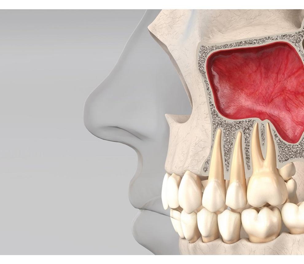
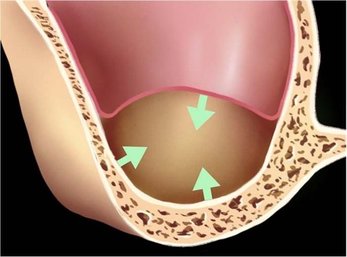
Anatomical variations of the maxillary sinus are common and may influence surgical outcomes. The presence of sinus septa, variations in membrane thickness, mucosal thickening, accessory ostia, and differences in sinus size and morphology can increase the difficulty of sinus augmentation procedures. Among these factors, sinus septa and thin Schneiderian membranes are particularly associated with a higher risk of membrane perforation during sinus floor elevation. Therefore, thorough preoperative assessment using Cone-Beam Computed Tomography (CBCT) is recommended to identify anatomical variations, facilitate surgical planning, and minimize complications during implant rehabilitation of the posterior maxilla.
3. Materials and Methods
3.1. Search strategy
A comprehensive electronic literature search was conducted using:
PubMed Google Scholar Cochrane Library Medline EBSCO Host
Studies published between 2012–2024 related to sinus-lift procedures and implant rehabilitation were included.
3.2. Inclusion criteria
Human clinical studies related to sinus-lift procedures. Studies evaluating implant survival and sinus augmentation outcomes. English-language articles. Studies involving posterior maxillary implant rehabilitation.
3.3. Exclusion criteria
Case reports with inadequate data. Animal studies. Review articles. Studies lacking clinical outcome evaluation.
3.4. Study selection
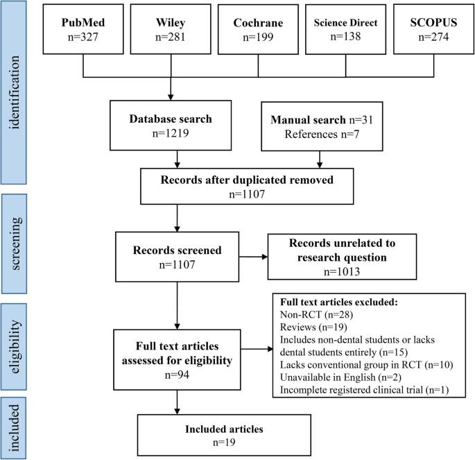
A total of 1037 studies were identified. After duplicate removal and screening, 19 studies fulfilled the eligibility criteria and were included in the systematic review.
4. Techniques of Sinus-lift Procedures
4.1. Lateral window technique
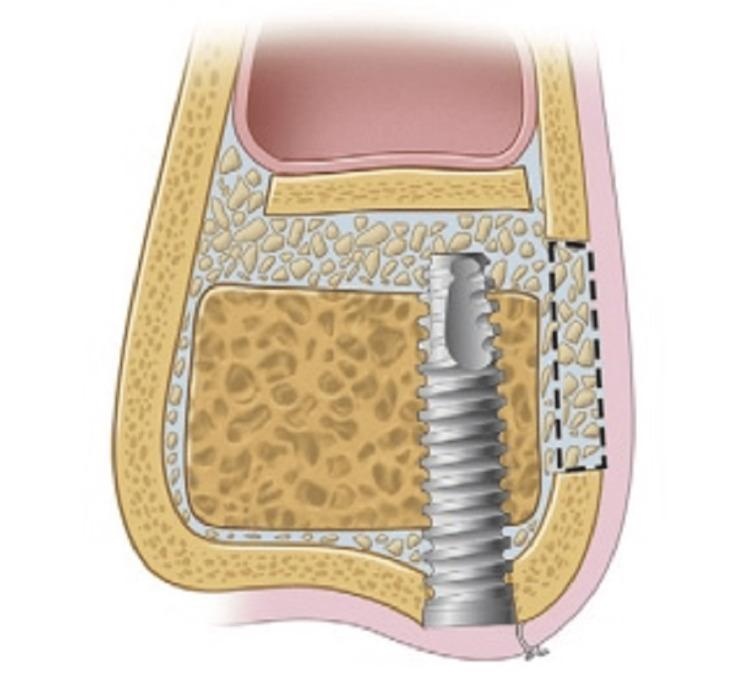
The lateral window technique involves reflection of a mucoperiosteal flap followed by creation of a bony window on the lateral wall of the maxillary sinus. The Schneiderian membrane is elevated carefully and graft material is placed beneath the membrane.
4.1.1. Advantages
Direct visualization of sinus membrane. Greater bone augmentation capability. Suitable for severe posterior maxillary atrophy.
4.1.2. Disadvantages
Higher surgical morbidity. Increased postoperative discomfort. Membrane perforation risk.
4.2. Crestal (transalveolar) technique
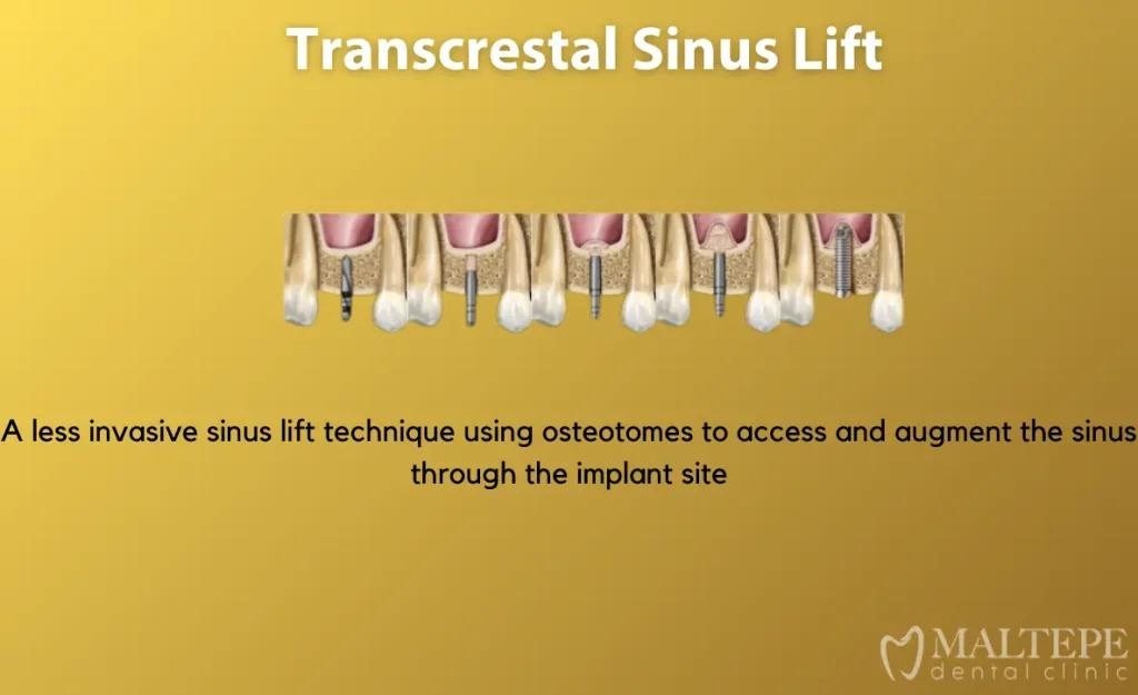
The crestal sinus-lift approach is minimally invasive and performed through the alveolar crest using osteotomes or specialized drills.
4.2.1. Advantages
Less invasive. Reduced postoperative morbidity. Shorter healing period.
4.2.2. Disadvantages
Limited augmentation height. Reduced visibility. Technique sensitive.
4.3. Hydraulic sinus lift technique
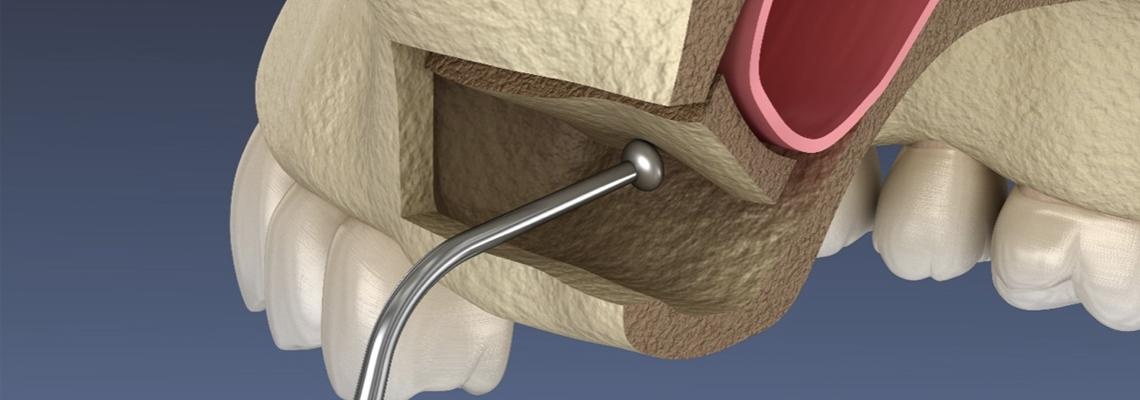
Hydraulic sinus lift procedures use hydraulic pressure to detach and elevate the Schneiderian membrane through a crestal approach. This method reduces surgical trauma and improves patient comfort while maintaining favorable clinical outcomes.
4.4. Piezoelectric Surgery Technique
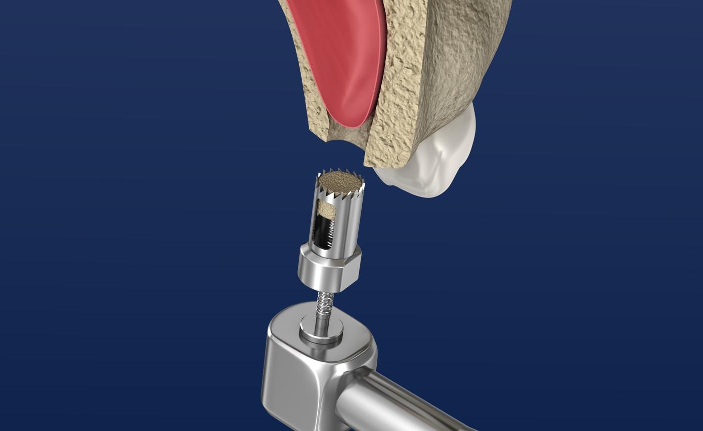
Piezoelectric surgery utilizes ultrasonic microvibrations for precise osteotomy preparation with minimal soft tissue injury.
Advantages include: Reduced membrane perforation. Precise bone cutting. Improved surgical safety.
4.5. Balloon-assisted sinus lift technique
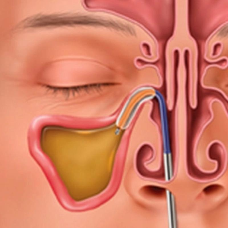
The balloon elevation technique involves insertion of a balloon device through the osteotomy site followed by gradual inflation to elevate the sinus membrane.This minimally invasive technique demonstrates reduced membrane perforation and improved patient comfort.
5. Results
Nineteen studies met eligibility criteria. Implant survival rates were consistently high across lateral window and crestal approaches. Minimally invasive procedures demonstrated reduced postoperative discomfort and favorable healing outcomes. Membrane perforation remained the most frequently reported complication. Nineteen studies evaluating various sinus augmentation procedures were included. Both lateral window and crestal approaches showed high implant survival rates and successful bone regeneration outcomes.
Minimally invasive techniques including hydraulic, balloon-assisted, and piezoelectric procedures demonstrated:
Reduced patient discomfort. Lower complication rates. Improved surgical precision. Faster healing periods.
Membrane perforation was identified as the most common intraoperative complication across all techniques.
|
Author (Year) |
Study Design |
Sample Size |
Sinus-Lift Technique |
Graft Material |
Follow-up Period |
Main Outcome |
|---|---|---|---|---|---|---|
|
Wallace et al. (2012) |
Prospective clinical study |
100 implants |
Lateral window |
Xenograft |
12 months |
High implant survival and bone gain |
|
Krasny et al. (2015) |
Clinical study |
42 patients |
Lateral window |
Allograft |
18 months |
Predictable sinus augmentation outcomes |
|
Rizzo et al. (2017) |
Retrospective study |
60 implants |
Crestal approach |
Xenograft |
24 months |
Reduced morbidity and favorable survival |
|
Helmy (2017) |
Clinical review |
35 patients |
Osteotome technique |
Mixed graft |
12 months |
Successful implant integration |
|
Alshamrani et al. (2023) |
Comparative study |
50 patients |
Piezoelectric surgery |
Xenograft |
24 months |
Reduced membrane perforation rate |
|
Pereira et al. (2024) |
Prospective study |
75 implants |
Hydraulic sinus lift |
Xenograft |
18 months |
Improved membrane elevation outcomes |
|
Alajami et al. (2024) |
Clinical study |
48 patients |
Balloon-assisted technique |
Alloplast |
12 months |
Minimally invasive and predictable results |
|
Technique |
Primary Indication |
Advantages |
Disadvantages |
Reported Clinical Outcome |
|
Lateral Window Technique |
Residual bone height <5 mm |
Direct visualization, large augmentation volume, suitable for severe atrophy |
More invasive, increased morbidity, longer healing period |
Excellent implant survival (>95%) |
|
Crestal (Transalveolar) Technique |
Residual bone height ≥5 mm |
Minimally invasive, reduced postoperative discomfort, shorter surgical time |
Limited augmentation height |
High implant survival (>94%) |
|
Osteotome-Mediated Technique |
Moderate sinus elevation |
Simultaneous implant placement, less invasive |
Risk of benign positional vertigo, limited elevation |
Favorable long-term outcomes |
|
Hydraulic Sinus Lift |
Moderate vertical augmentation |
Reduced membrane perforation, controlled elevation |
Technique sensitive |
High success and patient comfort |
|
Balloon-Assisted Technique |
Limited to moderate augmentation |
Atraumatic membrane elevation, minimally invasive |
Additional equipment required |
Predictable bone gain |
|
Piezoelectric Surgery |
Cases with complex sinus anatomy |
Precise osteotomy, reduced soft tissue injury, lower perforation risk |
Higher cost, increased operative time |
Excellent safety and implant survival |
CBCT-based preoperative planning is recommended for all sinus augmentation procedures to assess residual bone height, sinus anatomy, membrane thickness, and presence of sinus septa.
6. Discussion
When choosing the surgical technique, clinicians should assess the lateral wall thickness. A difference in the perforation rate was noted for a wall thickness measured at 6 mm coronal to Sinus pneumatization and ridge atrophy represent challenges to the successful rehabilitation of patients with posterior maxilla endosseous implants. Elevating the sinus comprises forming a “mucoperiosteal-alveolar pocket” above the maxillary floor and underneath the Schneiderian membrane to increase alveolar bone height and create a “confined space”.
Although both osteotome and lateral window procedures are safe and effective in atrophic posterior maxilla, residual bone height is crucial in determining the survival of these implants and sinus graft surgical decisions are majorly influenced by the RBH. With the improvement of implant surface modification and surgical equipment, the choice of MSFA approaches has been continuously updated and whether to choose immediate deferred placement of implants with anatomical variations is still controversial. No significant influence of RBH on the stability of the primary implant in atrophic sinus floor were reported, while initial RBH of less than 4 mm was reported to influence the success rates of implants inserted in combination with sinus floor elevation using osteotomes. Sinus elevation through the transcrestal window approach for a patient whose posterior maxilla had only 1–2 mm of residual alveolar bone was reported recently, and the incisions used in transcrestal window approach were shorter, compared with the lateral window approach, which could reduce discomfort of the patient after sinus elevation surgery. The evidence is insufficient and further long-term follow-up studies were needed. Meanwhile the transcrestal window approach requires a thorough assessment of the anatomy of sinus, elasticity, and deformation capacity of the Schneiderian membrane, the location of the intraosseous artery(which could be undetectable in CT/CBCT images), precise surgical approach, and In addition, the crestal approach was used to elevate the sinus floor of 27 patients with residual bone heights of 2–3 mm.Moreover, a recent study revealed that the RBH and vital bone formation were not statistically correlated.13 No significant differences in the amount of osteogenesis in sinuses classified as having an RBH ≤ 2 mm or >2 mm were observed the sinus floor. While other researchers also reported that lateral wall thickness had no effect on the perforation rate.
Despite the predictability of sinus lift procedures, intra-operative complications are common especially Schneiderian membrane perforation. Sinus compliance comprises the intrinsic potential of the sinus mucosa to resume its homeostatic status after the surgical trauma caused by sinus lifting. A higher rate of perforation is associated with a thinner membrane, possibly because the tensile capacity of a thicker membrane is significantly higher. Sinus augmentation surgery can be carried out on a 1–2 mm thick healthy and resilient membrane; however, for a thin membrane (<1 mm), a more cautious approach should be adopted. The Schneiderian membrane has the potential to thicken during inflammation, such as during odontogenic infections, especially apical infections.
Irritations, such as allergies, can also thicken the mucosa. However, sinus augmentation is not contraindicated by the presence of mild mucosal thickening or pseudo cysts in the absence of coexisting Sino nasal symptoms. However, with a deeper understanding of the maxillary sinus disease, some researchers formed different opinions, and diseases of the maxillary sinus should be diagnosed and managed carefully prior to sinus elevating surgery.
The stretching potential of Schneider’s membrane should be considered surgically and histologically. After lateral sinus floor elevation surgery, transient swelling of sinus membrane is observed, which reaching a peak value 7 days after surgery and completely resolves over months. The widths and contours of the sinus are closely related. For example, Wang et al. described the Palate-Nasal-Recess (PNR) as the intersection point of the two imaginary lines following the lower part of the lateral nasal wall and the palatal wall in the maxillary sinus, which would make elevation of the sinus membrane more complicated and enhance the risk of membrane perforation. Niu et al. considered the sinus width, sinus depth, the PNR, and angle A Simultaneously A flat or irregular maxillary sinus stretches more when lifted to the same height, which requires a better stretch potential of the Schneider membrane. For example, the presence of irregular bone walls, exostosis, and septa might contribute to the occurrence of sinus perforations. Perforation rates of MSFA when septa were present were much higher than in those without septa.
The lateral window technique remains the gold standard for severely atrophic posterior maxillae because it allows extensive augmentation and direct visualization. Crestal approaches are less invasive and suitable where residual bone height is adequate. Piezoelectric surgery improves surgical precision and decreases membrane injury. Hydraulic and balloon-assisted methods offer minimally invasive alternatives with encouraging outcomes. Selection of technique should be based on anatomy, augmentation requirements, clinician experience, and patient factors. Future developments include digital planning, navigation-guided surgery, tissue engineering, and growth-factor enhanced grafting.
Maxillary sinus augmentation has become a predictable and widely accepted procedure for implant rehabilitation in the atrophic posterior maxilla. The findings of this review indicate that various sinus-lift techniques provide favorable clinical outcomes when appropriately selected according to residual bone height, anatomical considerations, and treatment objectives.
The lateral window technique remains the gold standard for patients with severe posterior maxillary atrophy and limited residual bone height. This approach allows direct visualization of the Schneiderian membrane, facilitates substantial bone augmentation, and demonstrates high implant survival rates. However, it is associated with increased surgical complexity and postoperative morbidity compared with minimally invasive approaches ([Figure 4]).
The crestal or transalveolar sinus-lift technique is less invasive and is particularly suitable when adequate residual alveolar bone is present. Advantages include reduced surgical trauma, shorter operative time, and improved patient comfort. Nevertheless, the amount of achievable sinus elevation is limited when compared with the lateral window approach.
Recent advancements have led to the development of minimally invasive techniques, including hydraulic, balloon-assisted, and piezoelectric sinus augmentation procedures. Hydraulic sinus membrane elevation utilizes controlled fluid pressure to elevate the Schneiderian membrane, thereby reducing the risk of membrane perforation and improving patient comfort ([Figure 6]). Similarly, piezoelectric surgery enables precise osteotomy preparation with minimal trauma to adjacent soft tissues, resulting in enhanced surgical safety and reduced intraoperative complications.
Despite the high success rates reported for all sinus-lift procedures, membrane perforation remains the most frequently encountered complication. Other complications include bleeding, infection, graft displacement, and sinusitis. Careful preoperative assessment using Cone-Beam Computed Tomography (CBCT), meticulous surgical technique, and appropriate case selection are essential for minimizing these risks.
The reviewed literature demonstrates that implant survival rates following sinus augmentation are consistently high regardless of the technique employed. Therefore, the choice of procedure should be based on residual bone height, sinus anatomy, clinician experience, and patient-specific factors rather than a single universally applicable approach.
Future developments in sinus augmentation are expected to focus on digital treatment planning, computer-guided surgery, navigation-assisted implant placement, tissue engineering, and biologically active graft materials. Advances in regenerative medicine, including the use of platelet concentrates, stem cells, and growth factors, may further improve bone regeneration and treatment predictability while reducing patient morbidity.
Overall, sinus augmentation remains a reliable and effective procedure for increasing vertical bone height in the posterior maxilla, enabling successful implant placement and long-term functional rehabilitation.
Selection of the sinus-lift procedure depends on:
Residual bone height Sinus anatomy Presence of septa Membrane thickness Required augmentation Surgeon experience
The lateral window approach remains the gold standard in severely resorbed posterior maxilla because it allows greater augmentation and direct visualization. However, crestal techniques are associated with reduced surgical trauma and morbidity.
Recent advances in minimally invasive sinus augmentation techniques have significantly improved patient comfort and reduced complications.
7. Clinical Significance
Knowledge of sinus anatomy and available augmentation techniques allows clinicians to optimize implant success and minimize complications.
8. Limitations
Although sinus augmentation procedures have demonstrated high success rates, each technique possesses specific limitations that may influence treatment outcomes.
The lateral window technique, while considered the gold standard for severely atrophic posterior maxillae, is relatively invasive and associated with increased surgical time, postoperative discomfort, edema, and risk of Schneiderian membrane perforation. The procedure also requires greater surgical expertise and may result in prolonged healing periods.
The crestal (transalveolar) sinus-lift technique is less invasive but provides limited vertical bone augmentation. This technique is highly dependent on adequate residual bone height and may not be suitable for patients with severe maxillary atrophy. In addition, the risk of inadequate membrane elevation may compromise the amount of achievable bone gain.
The osteotome-mediated sinus floor elevation technique may cause patient discomfort due to malleting forces. Postoperative complications such as benign paroxysmal positional vertigo (BPPV), dizziness, and patient anxiety have been reported. Furthermore, the amount of sinus elevation achievable with this method is limited.
The hydraulic sinus-lift technique offers controlled membrane elevation; however, it is technique-sensitive and requires specialized equipment. Excessive hydraulic pressure may lead to membrane perforation if not carefully controlled.
The balloon-assisted sinus-lift technique provides atraumatic membrane elevation but increases treatment cost because of additional instruments and materials. In cases of complex sinus anatomy or the presence of septa, balloon placement may be challenging.
The piezoelectric sinus-lift technique significantly reduces the risk of soft tissue injury and membrane perforation; however, it requires specialized surgical devices, increased operator training, and longer operative times. The higher cost of equipment may limit its widespread use.
Overall, the choice of sinus augmentation technique should be based on residual bone height, sinus anatomy, patient-related factors, clinician experience, and available resources. No single technique is universally superior, and each approach presents specific advantages and limitations that must be carefully considered during treatment planning.
9. Conclusion
Sinus augmentation is the most common indication associated with implant placement in patients with severe edentulous maxilla. With the advancement of implant dentistry, there have been introductions of new techniques and grafting materials, which were aimed to improve the treatment outcomes of sinus augmentation. Several new concepts, such as the use of an angulated implant, zygomatic implant, or short implant, could provide another option for implantation in the posterior maxilla without the need for sinus augmentation, thus making treatment time shorter and reducing the rate of complications and the complexity of the treatment procedure.
Anatomical factors, including the remaining alveolar bone, maxillary sinus characteristics, the health condition of the Schneiderian membrane, and the lateral wall thickness, crucially affect successful lifting. The stretching potential affected by maxillary sinus characteristics and the health condition of Schneiderian membrane/sinus, as well as the residual bone height, influence clinical outcomes and the choice of surgical techniques, which should be taken into account by clinician.
Despite the reliability and efficiency of various sinus augmentation techniques, there is still a high rate of complications and complexity for such procedures. With the advances in technology and improvements in the design and manufacture of implants, some alternative concepts suggested implantation without sinus augmentation could be possible. The use of a tilted (angulated) implant in the posterior maxilla was suggested to avoid sinus augmentation. In this study, an evaluation was made to compare the efficiency between tilted and axial implants with no sinus grafting. After 5 years of follow-up, the implant success rate was 95.2% (survival: rate 100%) for the tilted implants and 91.3% (survival rate 96.5%) for the axial implants. The average marginal bone loss was 1.21 mm for the tilted implants and 0.92 mm for the axial ones.66 The concept of using a tilt implant was further enhanced. Trans sinus tilted implants, with the implant body inside the sinus, were utilized in the all-on-4 concept for complete edentulous maxilla patients. A survival rate of 96.4% was achieved at the implant level. The survival rate of prostheses was 100%. Sinusitis occurred in two patients (2.9%). The high survival rate and low complication rate suggest that trans-sinus implants could be an alternative solution to avoid sinus augmentation.
Zygomatic implants offer another option treatment modality to sinus augmentation. Almost similar to trans-sinus tilted implants, zygomatic implants are long implants that pass through the sinus or laterally to the sinus.68 The difference was the anchorage position. While the tip of a trans-sinus tilted implant is positioned in the bone between the anterior sinus wall and the nasal cortical bone,67 a zygomatic implant will anchor itself into the zygomatic process for stability. The use of a short implant (4–8 mm long) was also an interesting and straight forward alternate treatment modality for sinus augmentation followed by longer implant placement.
In a recent systematic review, there was further clarification of this concept. Eight RCTs from an initial search count of 851 titles were selected, and data extraction was performed. Both long-term follow-up (16–18 months) and short-term follow-up (8–9 months) study showed no significant differences when comparing implant survival rates. Most common complications were membrane perforations, and they were almost three times higher for longer implants in the augmented sinus compared to shorter implants. Morbidity, surgical time, and cost-effectiveness also showed more favourable data in the shorter implant group.
Sinus augmentation is a predictable procedure for rehabilitation of the posterior maxilla. Both lateral and crestal techniques demonstrate favorable outcomes. Minimally invasive techniques continue to expand treatment possibilities while reducing morbidity.
Sinus augmentation procedures are predictable and effective methods for implant rehabilitation in patients with inadequate posterior maxillary bone height. Both lateral window and crestal approaches demonstrate favorable clinical outcomes when proper diagnosis, treatment planning, and surgical technique are employed.
Minimally invasive approaches including hydraulic, balloon-assisted, and piezoelectric sinus-lift procedures provide promising alternatives with reduced morbidity and improved patient comfort.
10. Ethical Approval
Not applicable.
11. Authors Contributions
Nishi Bhati: Conceptualization, data curation, formal analysis, investigation, methodology, project administration, writing – original draft, Gunjan Gupta: Conceptualization, formal analysis, investigation, project administration, supervision, visualization, Sachit Anand Arora: Conceptualization, formal analysis, investigation, project administration, software, supervision, Shivesh Kumar Mishra: Conceptualization, data curation, formal analysis, funding acquisition, investigation, supervision, Priyanka Tandon: Data curation, funding acquisition, investigation, project administration, resources, validation,
12. Conflict of Interest
The authors declare no conflict of interest.
13. Source of Funding
Nil.
References
- Wallace SS, Tarnow DP, Froum SJ, Cho SC, Zadeh HH, Stoupel J. Maxillary sinus elevation by lateral window approach: evolution of technology and technique. J Evid Based Dent Pract. 2012;3382(12):70030-1. [Google Scholar] [Crossref]
- Alshamrani AM, Mubarki M, Alsager AS, Alsharif HK, AlHumaidan SA, Al-Omar A. Maxillary Sinus Lift Procedures: An Overview of Current Techniques, Presurgical Evaluation, and Complications. Cureus. 2023;15(11):e49553. [Google Scholar] [Crossref]
- Helmy MA. Maxillary sinus lifting: review of the two main approaches. Glob J Otolaryngol. 2017;8(4). [Google Scholar] [Crossref]
- Krasny K, Krasny M KA. Two-stage closed sinus lift: a new surgical technique for maxillary sinus floor augmentation. Cell Tissue Bank. 2015;16(4):579-85. [Google Scholar] [Crossref]
- Rizzo R, Checchi V, Marsili F, Zani A, Incerti-Parenti S, Checchi L. A New Transalveolar Sinus Lift Procedure for Single Implant Placement: The Ebanist Technique. A Technical Description and Case Series. Open Dent J. 2017;11:187-99. [Google Scholar] [Crossref]
- Lloret AM, Roge CP. Maxillary sinus lift surgery: description and comparison of different techniques. Int J Dent Med. 2024;10(2):31-4. [Google Scholar] [Crossref]
- Pereira RS, Carvalho MVNB, Carvalho MVNB, Hochuli-Vieira E, Statkievicz C, Santos DLP. Histomorphometric and micro-CT evaluation of Cerabone and Bio-Oss in maxillary sinus lifting: A Randomized Clinical Trial. Med. 2024;60(11):1834. [Google Scholar] [Crossref]
- Alajami MM, Elsheikh HA, Abo El-Farag SA, Mansour NA. Antral membrane balloon technique versus Densah bur in crestal sinus lift with simultaneous implant placement: a randomized clinical trial. BMC Oral Health. 2024;24(1):916. [Google Scholar] [Crossref]
- Wallace SS, Tarnow DP, Froum SJ, Cho SC, Zadeh HH, Stoupel J. Maxillary sinus elevation by lateral window approach: evolution of technology and technique. J Evid Based Dent Pract. 2012;3382(12):70030-1. [Google Scholar] [Crossref]
- Pereira RDS DCMHVE, Statkievicz C, Pereira Santos DL, Augusto Neto RT. Histomorphometric and Micro-CT Evaluation of Cerabone and Bio-Oss in Maxillary Sinus Lifting: A Randomized Clinical Trial. Med (Kaunas). 2024;60(11):1834. [Google Scholar] [Crossref]
- Alajami MM, Elsheikh HA, Abo El-Farag SA, Mansour NA. Antral membrane balloon technique versus Densah bur in crestal sinus lift with simultaneous implant placement: a randomized clinical trial. BMC Oral Health. 2024;24(1):916. [Google Scholar] [Crossref]
- Lloret AM, Rogel CP. Maxillary Sinus Lift Surgery: “Description and Comparison of the Different Techniques” Clinical Case of Sinus Lift and Placement of Phibo TSH and TSA Implants. Int J of Dent Med. 2024. p. 31-39. [Google Scholar] [Crossref]
- Ashwati, Krishna BJ, Bhandari N, Soman M, Gunasekar AS. A Maxillary Sinus Lift Technique by using New Maintained Active Bioline Implants-The Case Report. Int Clinc Med Case Rep Jour. 2024;3(1):1-14. [Google Scholar]
- Sala YM, Lu H, Chrcanovic BR. Clinical Outcomes of Maxillary Sinus Floor Perforation by Dental Implants and Sinus Membrane Perforation during Sinus Augmentation: A Systematic Review and Meta-Analysis. J Clin Med. 2024;13(5):1253. [Google Scholar] [Crossref]
- Albadani MM, Elayah SA, Al-Wesabi MA, Al-Aroomi OA, Al Qadasy NE, Saleh H. A graftless maxillary sinus lifting approach with simultaneous dental implant placement: a prospective clinical study. BMC Oral Health. 2024;24(1):227. [Google Scholar] [Crossref]
- Alshamrani AM, Mubarki M, Alsager AS, Alsharif HK, AlHumaidan SA, Al-Omar A. Maxillary Sinus Lift Procedures: An Overview of Current Techniques, Presurgical Evaluation, and Complications. Cureus. 2023;15(11):e49553. [Google Scholar] [Crossref]
- Abstract
- 1. Introduction
- 2. Anatomy of Maxillary Sinus
- 3. Materials and Methods
- 4. Techniques of Sinus-lift Procedures
- 4.1. Lateral window technique
- 4.2. Crestal (transalveolar) technique
- 4.3. Hydraulic sinus lift technique
- 4.4. Piezoelectric Surgery Technique
- 4.5. Balloon-assisted sinus lift technique
- 5. Results
- 6. Discussion
- 7. Clinical Significance
- 8. Limitations
- 9. Conclusion
- 10. Ethical Approval
- 11. Authors Contributions
- 12. Conflict of Interest
- 13. Source of Funding
- References
Article Metrics
- Visibility 476 Views
- Downloads 366 Views
- DOI 10.18231/j.jicd.48250.1785126220
-
CrossMark

- Citation
- Received Date May 22, 2026
- Accepted Date June 25, 2026
- Publication Date July 27, 2026