Journal of International College of Dentists
Journal of International College of Dentists (JICD) is an open-access, peer-reviewed, Quarterly journal publishing under the auspices of the International College of Dentists. As soon as the Article is accepted for Publication, it will be placed in “Ahead of Print” on the journal page with the aim of rapid and better knowledge dissemination. In addition, the journal allows free access (Open Access) to its contents, which is likely to attract more readers and citations to articles published in journal. Manuscripts must be prepared under the “Uniform requirements” of the ...
Role of ultrasonography in orofacial region
Abstract
Introduction: Ultrasonography (USG) has emerged as a valuable diagnostic imaging modality in the orofacial region due to its non-invasive nature, absence of ionizing radiation, real-time imaging capability, and cost-effectiveness.
Aims and Objectives: This review aims to evaluate the role of ultrasonography in the diagnosis and management of orofacial conditions. A comprehensive literature search was performed using electronic databases including PubMed, Medline, and ScienceDirect to identify studies related to ultrasonographic applications in oral and maxillofacial imaging. The reviewed literature demonstrates the utility of ultrasonography in the assessment of salivary gland disorders, cervical lymphadenopathy, temporomandibular joint disorders, vascular anomalies, maxillofacial infections, facial swellings, and soft tissue lesions.
Methodology: High-resolution ultrasonography and Doppler imaging have improved diagnostic accuracy, enabling better characterization of lesion morphology and vascularity. Recent advances such as three-dimensional ultrasonography, elastography, point-of-care ultrasound, and artificial intelligence-assisted imaging have further expanded its clinical applications.
Conclusion: Although limitations exist in imaging structures obscured by bone and air, ultrasonography remains a reliable adjunctive diagnostic tool for evaluating superficial soft tissue pathologies in the orofacial region. Continued technological advancements are expected to enhance its diagnostic potential and broaden its role in oral and maxillofacial practice.
1. Introduction
Ultrasonography is a non-invasive diagnostic imaging technique that employs high-frequency sound waves (above 20 kHz) to generate real-time images of internal anatomical structures without the use of ionizing radiation.[1] When the sound waves encounter interfaces between tissues with different acoustic impedances, a portion of the waves is reflected back to the transducer while the reminder continues through the tissues. The returning echoes are converted back into electrical signals by the same piezoelectric crystals. These signals are then processed by the ultrasound system to determine the location and intensity of the reflecting structures, thereby producing real-time cross-sectional images of the internal anatomy.[2]
2. Discussion
2.1. Ultrasonography in lymph nodes
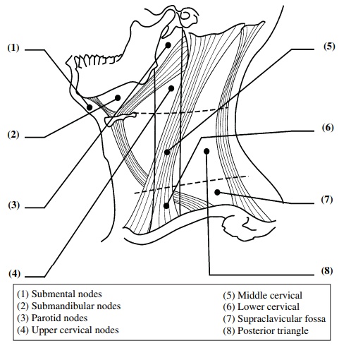
Cervical lymph nodes are commonly classified into seven levels based on the American Joint Committee on Cancer (AJCC) classification.[3] This classification has limitations in ultrasound examination, as certain nodal regions such as paratracheal nodes (Level VI) and upper mediastinal nodes (Level VII) are not accessible via ultrasound due to their deep anatomical location. An alternative classification system was proposed specifically for ultrasound evaluation by Hajek et al.[4] This system divides the cervical lymph nodes into eight regions, organized by their anatomical location within the neck ([Figure 1]). It was designed to promote a systematic approach to sonographic assessment of the cervical lymphatic chain.
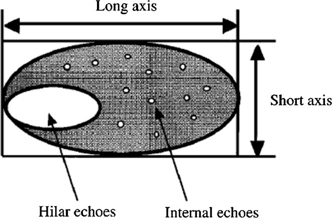
Ahuja et al mentioned that normal lymph nodes are usually hypoechoic when compared to the adjacent soft tissues, and tend to be oval or elongated in shape with a short axis to long axis ratio (S/L ratio) less than 0.5. The presence of an echogenic hilus (a hyperechoic linear structure continuous with adjacent soft tissues) is usually considered as a sign of benignity. Ultrasound findings evaluated in various studies mentioned that the longest dimension of a lymph node is referred to as the long axis, while the maximum perpendicular distance to the long axis is defined as the short axis ([Figure 2]). A homogeneous echogenic area within the lymph node, known as the hilar echo, represents the fatty tissue surrounding the hilum. The echoes of the parenchyma were defined as internal echoes which is helpful in guiding needle for FNAC. These internal echoes provide valuable information about the structural characteristics and homogeneity of the parenchyma, enabling differentiation between normal and pathological tissues.
In cases of lymphadenitis, affected lymph nodes typically appear enlarged, with an axial diameter exceeding 10 mm, and present with an ovoid to round configuration. Nonspecific inflammatory lymph nodes generally exhibit well-defined borders, but the hilum is often not visualized on ultrasound. In contrast, metastatic lymph nodes tend to show notable increases in size, a more rounded morphology, and internal heterogeneity. These changes are attributed to factors such as tumor necrosis, keratinization, or cystic degeneration within the metastatic deposit, all of which can be detected through ultrasonographic imaging.[5]
The lymph node status is a critical prognostic indicator in head and neck tumors and plays a pivotal role in guiding the treatment strategy. The presence, size, number, and characteristics of involved lymph nodes significantly influence both staging and overall prognosis, making accurate assessment essential for effective clinical decision-making.
Nodal size alone is not a reliable diagnostic criterion, as micro-metastases can occur in small nodes, and inflammatory conditions may cause benign enlargement. Study done on 142 subjects (702 lymph nodes) mentioned that besides nodal size, other various pathologies like distribution of lymphadenopathy, echogenicity, calcification, distal enhancement, intranodal cystic necrosis, matting and soft tissue oedema are also useful in further differentiating the malignant from benign lymph nodes.[6]
A criteria was proposed to diagnose the lymph nodes as benign or malignant:
A lymph node with hilar but no definite internal echoes is defined as benign. A lymph node measuring 10 mm or more in the short axis is defined as malignant. A lymph node with an L/S ratio of 3.5 or more is considered benign. A lymph node which cannot be associated to categories 1–4 is considered to be questionable.[7]
2.2. Ultrasonography of floor of the mouth
The floor of the mouth represents a complex anatomical region bounded superiorly by the ventral surface of the tongue and inferiorly by the mylohyoid and geniohyoid muscles.
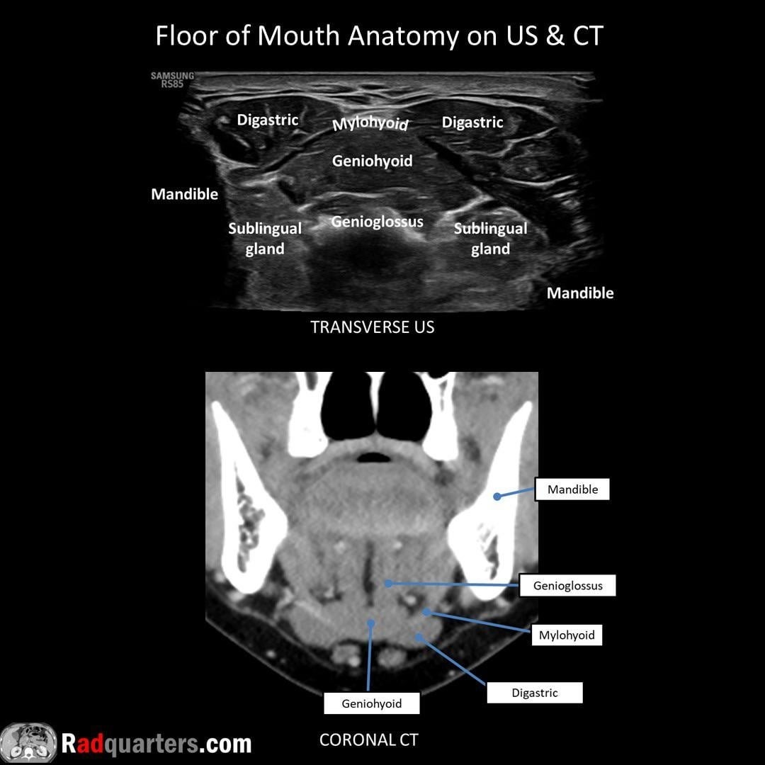
Ultrasonographic assessment of the floor of the mouth can be performed using two principal approaches. The most commonly employed technique, transcutaneous approach involves extraoral scanning through the submental region using high-frequency linear transducers, typically operating at frequencies greater than 9 MHz, which provides spatial resolution for superficial soft tissue structures ([Figure 3]).[8] This approach provides an excellent overview of the mylohyoid muscle, geniohyoid muscle, anterior belly of the digastric muscles, sublingual glands, submandibular glands and ducts, lingual musculature, and adjacent soft tissues. It is useful for evaluating inflammatory conditions, salivary gland pathologies, ductal calculi, cystic lesions, abscesses, vascular malformations, and tumors involving the floor of the mouth.
The intraoral approach involves placing a small high-frequency linear or hockey-stick transducer directly within the oral cavity over the floor of the mouth. This technique provides superior spatial resolution because the transducer is positioned in close proximity to the structures of interest, minimizing attenuation and improving image quality. The intraoral approach is particularly valuable for assessing superficial lesions, the sublingual space, the anterior portion of the submandibular (Wharton's) duct, small salivary calculi, mucoceles, ranulas, and other localized lesions that may not be adequately visualized using the transcutaneous technique.
2.3. Ranulas
Ranulas are mucus extravasation pseudocysts that arise predominantly from the sublingual gland due to ductal disruption or obstruction, resulting in the accumulation of saliva within the surrounding soft tissues. Clinically, they most commonly present as unilateral, fluctuant swellings in the floor of the mouth. A distinctive variant, known as the plunging or diving ranula, develops when mucin extends through a congenital or acquired defect in the mylohyoid muscle, allowing the lesion to spread into the submandibular or cervical spaces.
Sonographically, ranulas typically appear as well-circumscribed, thin-walled, anechoic or hypoechoic cystic lesions with posterior acoustic enhancement, closely resembling simple cysts ([Figure 4]). Suresh and Vora et al. in their studies demonstrated that ultrasonography can accurately identify ranulas and differentiate them from other cystic and solid lesions of the oral cavity.[9]
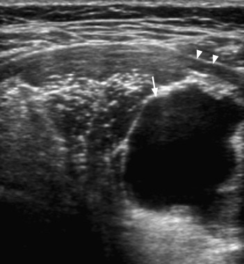
In cases of plunging ranulas, ultrasonography characteristically demonstrate a larger cystic component extending into the submandibular region with a tapering communication toward the sublingual space, commonly referred to as the “tail sign” or “beak sign”, which was highly specific imaging characteristic identified by Davison et al. and Zhi et al.[10]
2.4. Hemangioma of tongue
Hemangiomas are among the most common benign vascular tumors of infancy and childhood and frequently involve the head and neck region, including the oral cavity. Depending on their size, depth, and location, tongue hemangiomas may result in functional disturbances such as difficulties in speech, mastication, swallowing, and airway maintenance, in addition to causing significant aesthetic concerns.
Ultrasonography along with color Doppler imaging helps in evaluation of tongue hemangiomas, vascular architecture, and hemodynamic characteristics. Sonographically, hemangiomas typically appear as well-defined, compressible, hypoechoic soft tissue masses. Dubois et al. reported that hemangiomas characteristically exhibit marked internal blood flow and well-defined margins on Doppler ultrasonography, which facilitate their differentiation from other soft tissue lesions like lymphatic malformations, which generally demonstrate little or no internal vascularity.[11]
The sonographic appearance of hemangiomas may vary according to the stage of lesion development. Proliferative hemangiomas typically present as relatively homogeneous hypoechoic lesions with prominent vascular signals, whereas involuting lesions gradually become more heterogeneous and demonstrate reduced vascularity due to progressive fibrofatty replacement.[12] They also exhibit areas of fatty infiltration, fibrosis, or thrombosis, resulting in heterogeneous echotexture and variable Doppler findings.[13]
2.5. Ultrasonography in salivary gland disorders
The salivary glands are among the most accessible structures in the maxillofacial region for ultrasonographic examination because of their superficial anatomical location. The superficial lobe of the parotid gland and the submandibular gland can be readily visualized using high-frequency linear transducers, whereas evaluation of the deep lobe of the parotid gland remains relatively challenging because the mandibular ramus limits transmission of ultrasound waves.
2.5.1. Parotitis
Parotitis is one of the most common causes of parotid gland enlargement and may occur secondary to viral, bacterial, autoimmune, or obstructive etiologies. The affected glands typically appear enlarged and demonstrate multiple small, round hypoechoic areas measuring approximately 2–4 mm in diameter, corresponding to peripheral sialectasis, acinar destruction, and lymphocytic infiltration within the glandular parenchyma.[14]
2.5.2. Sialolithiasis
The submandibular gland is frequently involved because of long and tortuous course of Wharton’s duct, narrow lumen, and salivary flow against gravity. Obstruction of salivary outflow by calculi results in recurrent pain, swelling during meals, secondary infections, and chronic inflammatory changes within the gland.
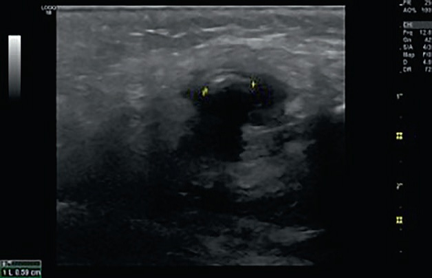
High-frequency transducers ranging from 7.5–12 MHz detect both calculi and associated glandular abnormalities with good spatial resolution for evaluating the ductal system and glandular parenchyma. Sialoliths appear as hyperechoic foci associated with posterior acoustic shadowing. Smaller or poorly calcified stones can be identified indirectly through associated findings such as ductal dilatation, gland enlargement, altered echotexture, and surrounding inflammatory changes.
One of the major advantages of ultrasonography is its ability to identify both radiopaque ([Figure 5]) and radiolucent calculi. Yoshimura et al. demonstrated that ultrasonography could reliably detect salivary stones that were not visible on conventional radiographs and simultaneously providing accurate stone size, location, ductal obstruction, and secondary glandular alterations.[15]
2.6. Salivary gland tumors
Salivary gland tumors comprise a diverse group of benign and malignant neoplasms with varying biological behavior and prognostic outcomes. The most common malignant salivary gland neoplasms include mucoepidermoid carcinoma, adenoid cystic carcinoma, acinic cell carcinoma, and adenocarcinoma.
Normal salivary gland parenchyma typically demonstrates a homogeneous echogenic appearance, allowing intraglandular lesions to be readily identified as focal abnormalities. Most salivary gland tumors appear as hypoechoic masses relative to the surrounding glandular tissue. Benign tumors generally appear as well-defined, homogeneous, smoothly marginated masses without associated cervical lymphadenopathy, whereas malignant tumors often demonstrate irregular or poorly defined borders, heterogeneous internal echotexture, infiltrative growth patterns, and associated metastatic cervical lymph nodeslthough these imaging features are not pathognomonic, they provide important clues regarding tumor behavior and help determine the need for further investigation.
Utrasonography can also be useful in evaluating specific tumor subtypes. Li et al. found that ultrasonographic findings closely correlated with CT characteristics regarding lesion borders and internal architecture.[16] These findings suggest that ultrasonography can provide substantial preoperative information while avoiding radiation exposure and reducing healthcare costs. Ishii et al. compared ultrasonographic findings with histopathological examination of surgically excised palatal tumors and concluded that ultrasonography was particularly valuable in the preoperative assessment of lesions measuring less than 3 cm in diameter.[17] The ability to determine lesion extent, depth of invasion, and internal characteristics before surgery assists clinicians in selecting the most appropriate surgical approach and minimizing operative morbidity.
2.7. Temporomandibular joint
Temporomandibular disorders (TMDs) comprise a heterogeneous group of musculoskeletal and neuromuscular conditions affecting the temporomandibular joint (TMJ), masticatory muscles, and associated structures. These disorders commonly present with pain, joint sounds such as clicking or crepitation, restricted mandibular movements, and functional impairment during mastication, speech, and swallowing.
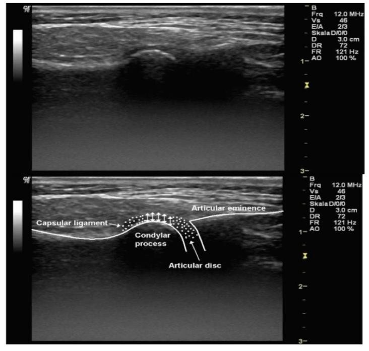
The temporomandibular joint is a superficial synovial articulation formed between the mandibular condyle and the glenoid fossa of the temporal bone. For ultrasonographic evaluation, high-frequency linear transducers, exceeding 12 MHz should be used which provide excellent spatial resolution for assessing superficial anatomical structures.[18] In ultrasonographic examination, the mandibular condyle and articular eminence are visualized as hyperechoic cortical outlines, whereas the articular disc appears as a thin hypo to isoechoic band interposed between the condyle and temporal bone. The joint capsule can also be identified as a hyperechoic structure surrounding the joint space ([Figure 6]).[19]
Among the various TMJ pathologies, internal derangement remains one of the most frequently encountered disorders. It commonly manifests as joint clicking, intermittent locking, pain, and restriction of mandibular movement. Studies by Bas et al. and Friedman et al. reported a sensitivity of 79% and 100% respectively in diagnosing temporomandibular joint disorders.[20][21]
The introduction of high-resolution ultrasonography (HR-US) has further enhanced diagnostic capabilities in TMJ imaging. Byahatti et al. observed that disc displacement is often more readily visualized in the closed-mouth position than in the open-mouth position. This phenomenon may be explained by medial displacement of the articular disc during mandibular opening, which can limit ultrasound beam propagation because of interference from surrounding osseous structures.[22]
Beyond internal derangement, ultrasonography has also contributed to the evaluation of normal joint biomechanics. Landes et al. investigated condylar translation and lateral joint space dimensions in asymptomatic individuals and demonstrated variations according to skeletal malocclusion patterns.[23]
3. Conclusion
Ultrasonography has become an indispensable imaging modality in the evaluation of diseases affecting the orofacial region owing to its absence of ionizing radiation, real-time imaging capability and wide availability. Its usefulness extends across a broad spectrum of conditions, including cervical lymphadenopathy, lesions of the floor of the mouth, salivary gland disorders, vascular anomalies, and temporomandibular joint pathologies, where it provides valuable diagnostic information and facilitates clinical decision-making. The advent of high-resolution and Doppler ultrasonography has further improved lesion characterization, assessment of vascularity, and guidance for interventional procedures. Furthermore, the growing adoption of ultrasound-guided fine-needle aspiration cytology (US-FNAC) and other image-guided interventions in oral and maxillofacial practice has enhanced diagnostic precision, improved sampling adequacy, and facilitated minimally invasive evaluation of salivary gland lesions and cervical lymphadenopathy, thereby contributing to more accurate treatment planning and reducing unnecessary surgical procedures.[24] As technological advancements continue and clinician expertise expands, ultrasonography can play an increasingly significant role in the accurate diagnosis, precise treatment planning, and follow-up of maxillofacial disorders.
4. Source of Funding
None.
5. Conflict of Interest
None.
References
- Rumack CM, Wilson SR, Charboneau JW, Levine D. Diagnostic Ultrasound. 5th ed. Philadelphia. 2018. [Google Scholar]
- Szabo TL. Diagnostic ultrasound imaging: inside out. Academic press. Academic press; 2013. [Google Scholar]
- Ying M, Ahuja A. Sonography of neck lymph nodes. Part I: normal lymph nodes. Clin Radiol. 2003;58(5):351-8. [Google Scholar] [Crossref]
- Hajek PC, Salomonowitz E, Turk R, Tscholakoff D, Kumpan W, Czembirek H. Lymph nodes of the neck: evaluation with US. Radiol. 1986;158(3):739-42. [Google Scholar] [Crossref]
- Evirgen Ş KK. Review on the applications of ultrasonography in dentomaxillofacial region. World J Radiol. 2016;8(1):50-8. [Google Scholar] [Crossref]
- Ahuja AT, Ying M, Ho SY, Antonio G, Lee YP, King AD. Ultrasound of malignant cervical lymph nodes. Cancer Imaging. 2008;8(1):48-56. [Google Scholar] [Crossref]
- Rama Mohan K, Koteswara Rao N, Leela Krishna G, Santosh Kumar V, Ranganath N, Vijaya Lakshmi U. Role of ultrasonography in oral and maxillofacial surgery: a review of literature. J Maxillofac Oral Surg. 2015;14(2):162-70. [Google Scholar] [Crossref]
- La'porte SJ, Juttla JK, Lingam RK. Imaging the floor of the mouth and the sublingual space. Radiographics. 2011;31(5):1215-30. [Google Scholar] [Crossref]
- Suresh BV, Vora SK. Huge plunging ranula. J Maxillofac Oral Surg. 2012;11(4):487-90. [Google Scholar] [Crossref]
- Davison MJ, Morton RP, McIvor NP. Plunging ranula: clinical observations. Head Neck. 1998;20(1):63-8. [Google Scholar] [Crossref]
- Dubois J, Garel L, Abela A, Laberge L, Yazbeck S. Lymphangiomas in children: percutaneous sclerotherapy with an alcoholic solution of zein. Radiol. 1997;204(3):651-4. [Google Scholar] [Crossref]
- Richter GT, Friedman AB. Hemangiomas and vascular malformations: current theory and management. Int J Pediatr. 2012;2012:1-10. [Google Scholar] [Crossref]
- Gibson CR, Barnacle AM. Vascular anomalies: special considerations in children. CVIR Endovasc. 2020;3(1):60. [Google Scholar] [Crossref]
- Baurmash HD. Chronic recurrent parotitis: a closer look at its origin, diagnosis, and management. J Oral Maxillofac Surg. 2004;62(8):1010-8. [Google Scholar] [Crossref]
- Yoshimura Y, Inoue Y, Odagawa T. Sonographic examination of sialolithiasis. J Oral Maxillofac Surg. 1989;47(9):907-12. [Google Scholar] [Crossref]
- Li J, Gong X, Xiong P, Xu Q, Liu Y, Chen Y, et al. Ultrasound and computed tomography features of primary acinic cell carcinoma in the parotid gland: a retrospective study. Eur J Radiol. 2014;83(7):1152-6. [Google Scholar] [Crossref]
- Ishii J, Nagasawa H, Wadamori T, Yamashiro M, Ishikawa H, Yamada T. Ultrasonography in the diagnosis of palatal tumors. Oral Surg Oral Med Oral Pathol Oral Radiol Endod. 1999;87(1):39-43. [Google Scholar] [Crossref]
- Kundu H, Basavaraj P, Kote S, Singla A, Singh S. Assessment of TMJ Disorders Using Ultrasonography as a Diagnostic Tool: A Review. J Clin Diagn Res. 2013;7(12):3116-20. [Google Scholar] [Crossref]
- Maranini B, Ciancio G, Mandrioli S GM, Govoni M. The Role of Ultrasound in Temporomandibular Joint Disorders: An Update and Future Perspectives. Front Med (Lausanne). 2022;9:926573. [Google Scholar] [Crossref]
- Bas B YNGE, Akan H. Diagnostic value of ultrasonography in temporomandibular disorders. J Oral Maxillofac Surg. 2011;69(5):1304-10. [Google Scholar] [Crossref]
- Friedman SN, Grushka M, Beituni HK, Rehman M, Bressler HB, Friedman L. Advanced Ultrasound Screening for Temporomandibular Joint (TMJ) Internal Derangement. Radiol Res Pract. 2020;2020:1-10. [Google Scholar] [Crossref]
- Byahatti SM, Ramamurthy BR, Mubeen M, Agnihothri PG. Assessment of diagnostic accuracy of high-resolution ultrasonography in determination of temporomandibular joint internal derangement. Indian J Dent Res. 2010;21(2):189-94. [Google Scholar] [Crossref]
- Landes CA, Goral WA, Sader R, Mack MG. 3-D sonography for diagnosis of disk dislocation of the temporomandibular joint compared with MRI. Ultrasound Med Biol. 2006;32(5):633-9. [Google Scholar] [Crossref]
- Kumar A, Khan J, Kaur G, Khalilullah M, Saha AK, Kumar A. Ultrasonography-Guided Fine Needle Aspiration Cytology Versus Histopathology: Diagnostic Consistency in Salivary Gland Neoplasms. Cureus. 2024;16(9):e69552. [Google Scholar] [Crossref]
- Abstract
- 1. Introduction
- 2. Discussion
- 2.1. Ultrasonography in lymph nodes
- 2.2. Ultrasonography of floor of the mouth
- 2.3. Ranulas
- 2.4. Hemangioma of tongue
- 2.5. Ultrasonography in salivary gland disorders
- 2.6. Salivary gland tumors
- 2.7. Temporomandibular joint
- 3. Conclusion
- 4. Source of Funding
- 5. Conflict of Interest
- References
Article Metrics
- Visibility 386 Views
- Downloads 382 Views
- DOI 10.18231/j.jicd.36277.1785127930
-
CrossMark

- Citation
- Received Date June 05, 2026
- Accepted Date July 10, 2026
- Publication Date July 27, 2026