Journal of International College of Dentists
Journal of International College of Dentists (JICD) is an open-access, peer-reviewed, Quarterly journal publishing under the auspices of the International College of Dentists. As soon as the Article is accepted for Publication, it will be placed in “Ahead of Print” on the journal page with the aim of rapid and better knowledge dissemination. In addition, the journal allows free access (Open Access) to its contents, which is likely to attract more readers and citations to articles published in journal. Manuscripts must be prepared under the “Uniform requirements” of the ...
Evaluation of the accuracy of methods for determining the working length of root canals in primary molars: A comparative clinical trial
Abstract
Introduction: The literature presents ongoing debate regarding the accuracy of various Working Length Determination (WLD) techniques in pediatric endodontic treatment. While some authors suggest that digital radiography may offer reliability comparable to—or even surpassing—that of conventional radiography, others report superior accuracy of electronic apex locators with conventional radiographic methods.
Aim: To evaluate the accuracy of Conventional Radiography (CR), Digital Radiovisiography (DR), and an Electronic Apex Locator (EAL) in WLD of root canals in primary molar teeth, with comparisons made against the Actual Root Canal Length (ARCL).
Materials and Methods: A total of 40 primary molars indicated for extraction were selected and the working length was determined using CR, DR, and an EAL, techniques. Following, these teeth were extracted and the ARCL was measured using Stereomicroscope. The obtained data was subjected to statistical analysis using One-way ANOVA and Chi-Square Test.
Results: In both maxillary and mandibular molars, working length obtained via CR and DR were significantly greater than those obtained using ARCL. Conversely, EAL measurements demonstrated close approximation to ARCL values. These variations were statistically significant (p ≤ 0.001).
Conclusion: The application of EAL demonstrated superior accuracy in determining the working length of root canals when compared to CR and DR, exhibiting a high degree of concordance with the ARCL.
1. Introduction
Accurate determination of the working length is essential for the success of pediatric endodontic therapy. Establishing a working length beyond the apical foramen may result in apical perforation, potentially injuring the periapical tissues and the developing permanent tooth germ, as well as causing the extrusion of irrigants and obturation materials. Conversely, underestimation of the working length may lead to insufficient debridement, retention of necrotic pulp tissue, persistent postoperative pain, and under-obturation.[1] The anatomical configuration of root canals in primary teeth—particularly molars—is inherently variable and challenging to predict due to ongoing physiological root resorption. This process alters the morphology, dimensions, and spatial orientation of the root apex over time, complicating accurate localization of the apical terminus.[2]
Conventional Radiography (CR) has long served as the standard modality for determining the working length in primary teeth. Periapical radiographs play a vital role in diagnosis, enabling visualization and assessment of root canal morphology. However, acquiring high-quality radiographic images in pediatric patients presents considerable challenges. Factors such as anatomical superimposition, variability in apical foramen morphology, image distortion, difficulty in visualizing resorptive changes, and limited patient cooperation can compromise image quality and diagnostic accuracy. Inadequate radiographic techniques, influenced by the constrained dimensions of the pediatric oral cavity, oversized film holders, and incompatible film sizes, further impede precise localization of the root apex.[1][3] These limitations are particularly problematic in cases of root resorption, where the radiographic apex may not accurately correspond to the actual anatomical terminus.[4]
Advancements in imaging technology have introduced Digital Radiovisiography (DR) as a promising alternative for determining endodontic working length. DR enables real-time visualization of radiographic images on digital monitors, thus enhancing diagnostic capability and patient engagement. It offers superior accuracy compared to CR, reduces radiation exposure, expedites image acquisition, and allows for magnification of critical regions such as the apical area. These capabilities collectively improve diagnostic efficacy and clinical workflow by eliminating conventional film processing. DR also integrates computational tools for image capture, enhancement, display, and archiving [1][2]. However, its widespread adoption is tempered by the substantial cost associated with installation and maintenance.
Nonetheless, both CR and DR share inherent limitations due to the two-dimensional representation of three-dimensional anatomical structures. These methods cannot adequately depict the buccolingual aspects of the root canal system because of overlapping anatomical features such as dentin, cementum, cortical bone, and alveolar structures. Furthermore, lateral canals, accessory foramina, and apical constrictions often go undetected, while subtle or oblique patterns of root resorption may be indistinct radiographically. The determination of working length via radiographs is also subject to interpretive variability and may present misleading images—for instance, suggesting intracanal placement of endodontic instruments when they are, in fact, extra-canal.[5]
Electronic Apex Locators (EALs) have emerged as viable alternatives to radiographic techniques for the estimation of working length in primary teeth since their initial introduction. EALs define the working length by measuring the impedances with different frequencies between the endodontic file and the canal fluids. A key benefit of EALs is their ability to determine working length to the apical foramen rather than the radiographic apex, thereby offering enhanced anatomical accuracy.[2] The devices are especially advantageous in pediatric endodontics, as they provide a more rapid, comfortable, and objective assessment that remains accurate even in the presence of physiological root resorption.[1][2][3][6] Further, they notably reduce the exposure to ionizing radiation and decreased procedural time.[2] Additionally, EALs circumvent several limitations associated with radiographic imaging—such as image distortion, anatomical superimposition, and patient cooperation challenges—particularly in children who exhibit strong gag reflexes during intraoral radiographic procedures.
The literature presents ongoing debate regarding the accuracy of various working length determination (WLD) techniques in endodontic treatment. While some authors suggest that digital radiovisiography may offer reliability comparable to—or even surpassing—that of conventional radiography,[7] others report superior accuracy with conventional radiographic methods.[8][9] Reported precision levels further highlight this discrepancy, with studies indicating accuracy rates of 82% for CR and up to 95% for EALs.[5][10] Numerous clinical investigations have evaluated the performance of different EALs in determining working length in primary teeth and demonstrate a broad range of reported accuracy, spanning from 64% to 96.7%.[5][11][12][13][14][15][16][17][18][19] These findings collectively reflect the variability in EAL performance and underscore the importance of continued research to clarify their clinical efficacy in primary dentition.
The majority of existing investigations are in-vitro, with relatively few in-vivo studies available specifically addressing primary teeth. Furthermore, these studies predominantly focused on single-rooted teeth, with minimal and uneven representation of primary molars and primarily conducted on teeth scheduled for extraction, introducing potential bias into their findings. Additionally, authors of a recently conducted meta-analysis highlighted the significant concerns regarding the methodological quality and heterogeneity of the included clinical trials.[19] Thus, more robust evidence is necessary to substantiate the accuracy of these devices in the context of primary dentition. Therefore, the purpose of the present study is to evaluate the accuracy of Conventional Radiography, Digital Radiography, and an Electronic Apex Locator in determining the working length of root canals in primary molar teeth, with comparisons made against the Actual Root Canal Length (ARCL), serving as the gold standard.
2. Materials and Methods
2.1. Ethical considerations
The protocol of this comparative research received approval from the Institutional Ethics Committee and was registered under clinical trials. The study adhered to the ethical principles outlined in the World Medical Association's Declaration of Helsinki. Additionally, the study followed the CONSORT guidelines for the design and reporting of clinical trials ([Figure 1]).
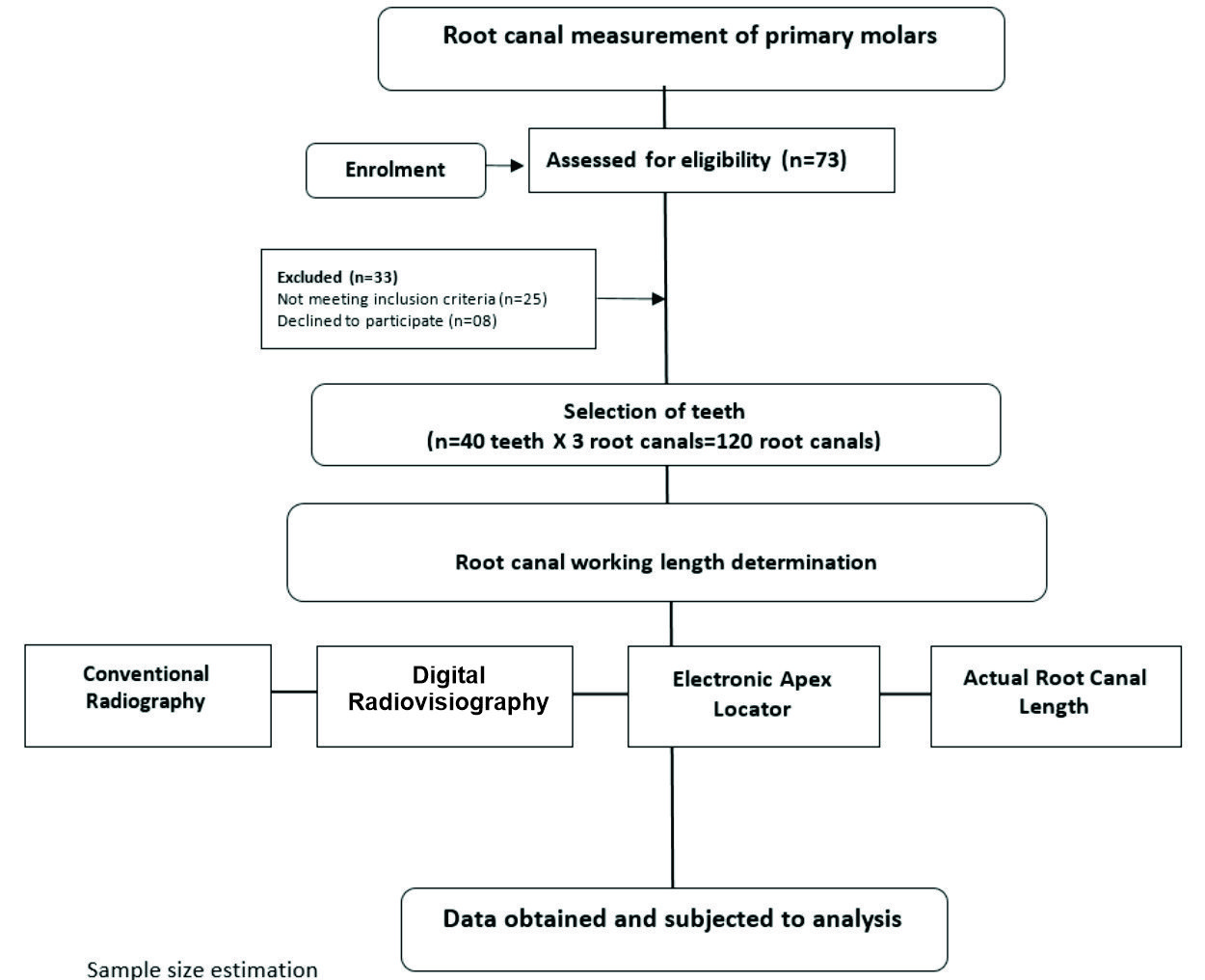
The sample size was determined using G*Power software (version 3.1.9.7; Heinrich Heine University, Düsseldorf, Germany). Drawing upon data from previous studies,[5][6] the mean difference between electronic and actual root canal lengths (measured in millimetres) was used to calculate the effect size, which was estimated at 0.18. With a significance level (α) of 5% and a statistical power of 95%, the required sample size was calculated to be 86 root canals. Accounting for a potential 10% attrition rate, the adjusted sample size increased to 96 root canals, corresponding to 32 teeth. To ensure balanced representation across all primary molar types, the final sample size was rounded up to 120 root canals canals (5 X 8 types of molars=40 teeth) ([Figure 1]).
2.2. Selection of study participants
Healthy, cooperative children aged 4 to 7 years participated in the study. Parents or legal guardians were thoroughly briefed on the study's objectives, clinical procedures, treatment outcomes, and the risks and benefits involved. Their informed written consent was obtained after all their questions were addressed. Additionally, children aged over 6 years provided their assent. Children with special healthcare needs, limited or no cooperative abilities, those needing sedation or general anaesthesia for behaviour management, and those allergic to local anaesthetics or other drugs were excluded,[17][20] Children with primary molars indicated for extraction due to dental mobility, over-retention, non-restorability, orthodontic treatment, extensive caries, unwillingness of parent to save the teeth in children aged 4–7 years were selected. Teeth with radiographic evidence of absence of root splits, cracks, furcation or root perforations, and abnormal root morphology (eg dilaceration), root fractures, advanced root resorption, pulpectomy and pulpotomy, calcified and obliterated root canals and more than 3 root canals were excluded,[1][21] A total of 73 teeth were screened, and 40 were selected.
2.3. Training and calibration
The co-investigators JC and KG underwent calibration to conduct CR and DR measurements, respectively. Working length measurements for ten radiographs of primary molars requiring pulpectomy were performed twice, with a seven-day interval between each measurement. All radiographs were obtained from the hospital database, ensuring that no unnecessary radiographic procedures were conducted on children. Co-investigator PK was calibrated to perform electronic measurements. For this purpose, working length determination using an EAL were performed on ten teeth undergoing pulpectomy, spaced seven days apart. Co-investigator GMD was calibrated to determine the ARCL using a stereomicroscope on eight primary molars. Additionally, a pilot study involving ten children was conducted to standardize measurement techniques, during which WLD was performed on fifteen primary molars. No significant adjustments were required. Children who participated in the calibration process or the pilot study were excluded from the final sample. The kappa test scores for investigators JC, KG, PK and GMD were 0.86, 0.93, 0.88 and 0.96, respectively.
2.4. Tooth preparation
The endodontic procedure commenced with drying of the injection site, followed by the application of a topical anesthetic and subsequent administration of local anesthesia. An access cavity was prepared in the selected primary molars. Coronal pulp tissue was removed using a spoon excavator, and the radicular pulp was extirpated with H-files (Mani, Inc., Tochigi, Japan). Residual pulpal tissue and dentinal debris were eliminated through copious irrigation with saline, and the canals were subsequently dried using sterile paper points.
Patency of the root canals was verified, and the canal orifices were enlarged using a coronal-flaring instrument (Endoflare®; Micromega, Geneva, Switzerland). Thereafter, the primary operator (GB) exited the operatory to ensure blinding, and the co-investigators (JC, KG, and PK) independently determined the working length using three distinct methods: conventional radiography, digital radiovisiography, and an electronic apex locator. The procedural steps employed for working length determination are detailed below.
2.5. Determination of working length of the root canals using conventional radiograph[22]
The distance from the cusp tip to the apex or the border of physiological/pathological root resorption was determined by superimposing an endodontic instrument (ISO #15 and #20 K files) over the preoperative radiograph, measured using a calibrated digital caliper. Subsequently, the rubber stopper on the files was adjusted to be 1-2mm short of these measurements. The files were then inserted into the root canals and a conventional radiograph was taken using the paralleling technique. Intraoral radiographs were captured using a dental X-ray unit (Confident Dental Equipment Ltd, New Life Radiology, Italy) operating at 60 kVp, 6 mA, 0.3 seconds, with exposure times of 200 ms for maxillary teeth and 120 ms for mandibular teeth. The distances between the source and the tooth, as well as the tooth and the films, were standardized using an X-ray positioner, which was consistently placed in the same position and adjusted for the size of the child's mouth for comfort. The radiograph was developed and fixed according to standardized protocols. On the second radiograph (working length radiograph), the actual working length measurements were determined in a dark room using an X-ray viewer and a 4X magnifying glass, and the measurements were recorded.
2.6. Determination of working length of the root canals using digital radiovisiography[23]
A digital radiovisiography device (Vista Scan Mini Plus, Dürr Dental; Bietigheim-Bissingen, Germany) with a "Size 0" phosphor plate sensor was used to obtain digital radiovisiograph. The radiograph was taken using a standardized paralleling technique with a sensor holder and circular collimation at a distance of 30 cm. Each tooth was imaged using an X-ray device (Confident Dental Equipment Ltd, New Life Radiology, Italy) operating at 70 kVp and 8 mA, with an image exposure time of 0.16 s. The sensor was scanned in the scanner to obtain digital images. The distance from the reference point on the crown to the end of the file, i.e., the radiographic apex, was measured on the computer monitor using integrated software (DBSWIN v 5.3.1, Dürr Dental).
2.7. Determination of working length of the root canals using electronic apex locator[12,23]
Electronic root canal length measurement was performed by placing the clip on the patient’s lip and inserting a 15/0.02 K-type file (Dentsply Maillefer, Baillagues, Switzerland) into the root canal until the EAL (Root ZX II; J. Morita, Tokyo, Japan) beeped and the panel indicated that the apex level had been reached. This measurement was deemed valid when the value remained stable for at least 5 seconds. The rubber stopper was adjusted to the reference point, the file was carefully removed from the canal, and the measurement was taken using calibrated digital calipers.
Following WLD with all the three methods, the tooth was extracted and the root surfaces were inspected for the presence of perforations or any other abnormalities that could have influenced the working length estimation.
|
Root Canals |
CR vs DR |
CR vs EAL |
CR vs ARCL |
DR vs EAL |
DR vs ARCL |
EAL vs ARCL |
|---|---|---|---|---|---|---|
|
Maxillary molars # |
||||||
|
Palatal |
<0.001* |
<0.001* |
<0.001* |
0.001* |
<0.001* |
<0.001* |
|
Mesiobuccal |
<0.001* |
<0.001* |
<0.001* |
<0.001* |
<0.001* |
<0.001* |
|
Distobuccal |
<0.001* |
<0.001* |
<0.001* |
<0.001* |
<0.001* |
<0.001* |
|
Mandibular molars # |
||||||
|
Distal |
<0.001* |
<0.001* |
<0.001* |
0.01* |
<0.001* |
<0.001* |
|
Mesiobuccal |
<0.001* |
<0.001* |
<0.001* |
<0.001* |
<0.001* |
0.04* |
|
Mesiolingual |
0.005* |
0.05* |
0.03* |
<0.001* |
0.05* |
0.05* |
2.8. Determination of actual root canal length using stereomicroscope[14]
Following extraction, the selected teeth samples were immediately immersed in a 0.9% saline solution. Within 24 hours, a cleaning protocol was implemented to remove soft debris, calculus, and stains using manual scrapers and ultrasonic scalers. Subsequently, the samples were stored in dark glass containers filled with a 1% thymol solution at room temperature for a week to ensure disinfection and maintain sample hydration, preventing dehydration-induced brittleness. Afterward, the samples underwent thorough rinsing with tap water for two hours to eliminate residual thymol.
The teeth samples, with a stabilized file in the canal, were meticulously sectioned longitudinally using a diamond bur mounted on a straight handpiece until the canal became visible. Careful removal of the thin dentin layer was performed to expose the canal while ensuring the instrument remained undamaged. Remaining traces of dentin were extracted with a probe, revealing the canal and the endodontic file positioned within it. This preparation enabled observation of the canal's topography and the spatial relationship between the file tip, apical constriction, and anatomical apex under a stereomicroscope.
Measurements of the root canal length were conducted using a stabilized K file (Dentsply Maillefer), adjusted to a coronal reference point with a rubber stopper. The file was inserted into each root canal until its tip was barely visible at the apical foramen. At 15x magnification, a Stereomicroscope (Leica Microsystems, Heerbrugg, Switzerland) captured photographic images of the samples. Using the integrated "straight-line measurement" feature, the ARCL was quantified by measuring from the rubber stopper's base to the file tip.
2.9. Evaluation of accuracy of methods of determining of working length of root canals[21,17]
For each root canal, the measurement procedure was performed three times. The average of these repeated measurements was then calculated to establish the working length of the root canal. For each method, the mean working length values were computed by subtracting 1 mm from the measured lengths. These adjusted mean values were subsequently compared with the ARCL (Gold Standard) to assess the accuracy of the WLD methods.
2.10. Blinding
The participants, their parents, outcome assessors, and data analysts were blinded to the method of working length determination.
2.11. Data analysis
Statistical Package for Social Sciences [SPSS] for Windows Version 22.0 Released 2013. Armonk, NY: IBM Corp., will be used to perform statistical analyses. Descriptive Statistics: Descriptive analysis of all the explanatory and outcome parameters was done using frequency and proportions for categorical variables, whereas in Mean±SD for continuous variables. Inferential Statistics: One-way ANOVA Test followed by Tukey's post hoc Test was used to compare the mean age, mean working length of different canals in maxillary and mandibular molars. Chi-Square Test was used to compare the gender distribution and teeth considered. The level of significance was set at p<0.05.
3. Results
The ages of the participating children ranged from 4–7 years, with a mean age of 5.46 ± 1.03. The involved a total of 40 teeth, which included 17 teeth from 12 male children and 23 teeth from 18 female children. The mean differences in relation to age and gender were not statistically significant. In maxillary molars, the mean root canal working length of the palatal root canal was recorded as 13.33 ± 0.19 mm using CR, 13.09 ± 0.08 mm using DR, 12.10 ± 0.29 mm with the EAL, and with 12.73 ± 0.12 mm of ARCL. Similarly, in mandibular molars, the distal root canal exhibited root canal working length of 13.32 ± 0.22 mm (CR), 13.05 ± 0.03 mm (DR), 12.09 ± 0.24 mm (EAL), and 12.88 ± 0.12 mm (ARCL). In both maxillary and mandibular molars, working length obtained via CR and DR were significantly greater than those obtained using ARCL. Conversely, EAL measurements demonstrated close approximation to ARCL values. These variations were statistically significant (p ≤ 0.001). A consistent pattern was also noted in the WLD of the maxillary mesiobuccal and distobuccal canals, as well as the mandibular mesiobuccal and mesiolingual canals. Furthermore, the root canal lengths measured using EAL were notably lower than those determined using CR and DR ([Figure 2]).
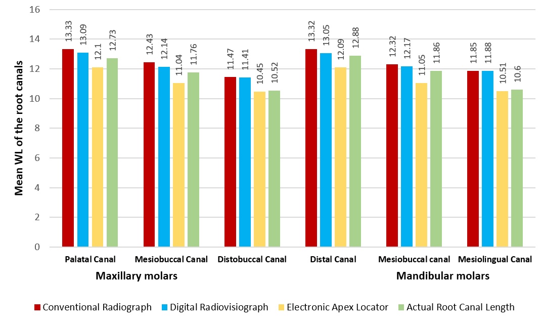
A significant association was observed between root canal lengths measured by EAL and ARCL in multiple comparison analyses ([Table 1]).
4. Discussion
Traditionally, radiographic imaging has been employed to visualize root canal anatomy, establish the working length, and assess the condition of apical tissues. In consistent with previous literature,[24][25] both radiographic methods (CR and DR) demonstrated a tendency to overestimate root canal length determination in comparison to EAL and ARCL. This discrepancy is primarily attributed to physiological and pathological root resorption in primary teeth, which predominantly occurs on the surfaces adjacent to the developing permanent successor tooth germ in buccolingual direction. These resorptive changes, particularly those involving oblique surfaces, are often not discernible on radiographic imaging.[26] As the resorptive process advances, the apical foramen undergoes progressive positional changes, often resulting in root canals that terminate at a considerable distance from the radiographic apex or designated apical reference point, thereby posing additional challenges to accurate working length determination. Thus, it is critical to note that radiographic assessments reflect the radiographic apex rather than the true anatomical location of the apical foramen.[4]
Moreover, anatomical variations—such as the apical foramen being located short of the radiographic apex on either the facial or lingual aspect of the root—pose significant challenges to the accurate radiographic determination of apical positioning. This complexity is further amplified in multirooted teeth, where identifying the precise apical termination is inherently more difficult than in single-rooted counterparts, potentially contributing to canal length overestimation when using radiographic techniques.[19]
The radiovisiography system used in the present study utilizes Photostimulable Phosphor (PSP) technology to record incident X-rays on the image receptor. The observed overestimation of root canal lengths may be attributed to the larger pixel size of the blue plates, as resolution is inversely proportional to pixel size. Furthermore, magnification errors may arise due to sensor positioning, despite the use of position-indicating devices, as the absence of a precise reference point compromises accuracy. Furthermore, existing evidence suggests that digital imaging systems are generally less effective in rendering radiopaque structures.[27] This limitation potentially hinders the precise localization of the file tip and, consequently, the accurate measurement of file/root length in PSP-derived images. Nevertheless, the practical limitations associated with solid-state sensors—such as a limited active imaging area, bulkier sensor design, and the presence of connecting cables—render them less suitable for use in pediatric dental settings.[2]
In the present study, the working length determined using the EAL was insignificantly shorter than the ARCL, a finding that corresponds with previous studies.[5][28] These authors reported that EAL accuracy is influenced by the diameter of the apical foramen, where wide apical openings—often resulting from physiological or pathological root resorption—along with flared canal morphologies, can lead to slight underestimation of the actual length. Additionally, in primary teeth, this discrepancy is likely due to the potential invagination of periapical soft tissues into the canal through lateral canals or enlarged apical foramina, which may contribute to underestimation of the working length.[26] However, the risk of underestimating the working length can be minimized through the use of pre-operative diagnostic radiographs.
Ideally, canal preparation should end at the cemento-dentinal junction or the apical constriction;[29] however, due to the resorptive processes inherent in primary dentition, a clear apical constriction may not always be identifiable.[30] However, clinically, several investigators have considered a variance of ±0.5 mm to ±1 mm between electronically derived and directly measured working lengths is generally deemed acceptable in primary teeth reinforcing the clinical reliability of EALs in pediatric endodontics.[4][11][15][31]
The present study demonstrated that, under in-vivo conditions, the EAL exhibited superior accuracy in root canal length determination compared to DR and CR. This finding is in line with previous clinical investigations that have consistently reported that EALs represent the most precise approach for determining the working length in primary teeth when compared to other methods.[3][14][21][31] Additionally, researchers have indicated a 95–96% accuracy rate in primary teeth when using EALs versus traditional CR and DR methods.[4][11][32] Supporting evidence from Khan et al. demonstrated that the Root ZX Mini apex locator achieved greater accuracy in identifying the true working length when compared to integrated endomotors with inbuilt apex locators and DR.[33] Likewise, Abdullah et al. confirmed that newer-generation EALs showed close agreement with DR measurements.[5]
Multiple factors can affect the accuracy of electronic root canal length measurements. A key anatomical variable is the frequent lateral displacement of the apical foramen, which often deviates from the apico-central axis. Additionally, buccolingual curvatures in the root canal may lead to discrepancies between electronic working length determinations and those obtained through radiographic techniques.[16] Physiological and pathological root resorption further complicate measurement accuracy. In cases of minimal resorptive defects, particularly on the buccal or lingual root surfaces, EALs typically produce readings shorter than those obtained radiographically, yet closer to the ARCL. When discrepancies between electronic working length and radiographic measurements are less than 2 mm, the EAL readings may be deemed more dependable. However, discrepancies exceeding 2 mm necessitate an additional working length radiograph to rule out potential canal perforation or device malfunction.[16] Nonetheless, despite challenges such as apical enlargement and resorptive changes, previous studies have confirmed the reliability of the electronic apex locator in primary teeth, owing to the preservation of the canal's conical taper.[4][34]
Although the present study demonstrated superior accuracy of the EAL compared to the two radiographic methods evaluated, contrasting findings have been reported in the literature. Katz et al. and Mente et al. reported no statistically significant differences EALs and CR in in-vitro assessments.[35][36] Similarly, Subramaniam et al. conducted an in-vitro investigation comparing the digital tactile sense technique, EAL, CR and DR methods with stereomicroscopy (considered the ARCL) to determine the working length in primary single-rooted teeth. Their results demonstrated no statistically significant differences among the techniques assessed.[37] Bodur et al. observed a statistically significant discrepancy between EAL-based measurements and ARCL in primary teeth, regardless of the presence of root resorption.[7] Rathore et al. and Chougule et al. reported no significant difference between working lengths derived using apex locators and radiographic methods.[13][26] Similarly, Martinez-Lozano et al. and Kqiku et al. concluded that no single method consistently achieved reliable results in actual working length determination, and that the accuracy of EALs did not exceed that of radiographic approaches.[38][39] Research conducted by Balaji et al. and Davalbhakta et al. also found no statistically significant difference in WLD accuracy between DR and EALs.[11][15] In line with these findings, Ghule et al. emphasized that EALs should not be considered the most dependable modality for working length assessment, particularly when compared to advanced imaging technologies such as Cone Beam Computed Tomography.[40] Other researchers have proposed that EALs function best as a complementary tool to radiographic techniques.[4][37]
The discrepancies observed across various studies may largely stem from methodological variations, particularly the in-vitro nature of certain investigations. Recreating the complex oral environment outside the oral cavity presents significant challenges, notably due to the absence of the periodontal ligament, which can compromise the accuracy of EALs. Additional contributing factors include small sample sizes, inconsistent tooth selection (e.g., with or without resorption), uneven representation of single- versus multirooted teeth, lack of comparison to ARCL, differing evaluation approaches (assessing the tooth as a whole versus individual root canals), and the use of various EAL models.[4][11][15][18] In contrast, the present study represented a large and evenly distributed sample of primary molars (multirooted teeth), with each root analysed individually to facilitate a more comprehensive evaluation of WLD across all selected methods. This approach addressed the limitations of previous studies, which were conducted on smaller sample sizes,[3][6][35] primarily focused on single-rooted teeth,[3][35] and considered only the total number of canals[4] rather than assessing each root separately. These rigorous methodological distinctions likely contributed to the differences in findings reported across studies.
This study, however, has certain limitations. The reference standard used was stereomicroscopic examination—an ex-vivo technique—which may introduce discrepancies when compared with in-vivo assessments. Hence, future research employing in-vivo gold standard methodologies, such as micro-computed tomography (Micro-CT), is recommended to validate and expand upon these findings. Furthermore, it is important to acknowledge that the intraoral nature of the radiographic technique may have introduced a degree of image distortion or reduced clarity due to angulation errors.
The findings of the present study substantiate the efficacy of EALs in accurately determining working length in primary teeth and advocate their integration into pediatric endodontic practice to optimize treatment protocols. Nonetheless, the indispensable role of an initial diagnostic radiograph must be emphasized. Radiographic evaluation remains essential for establishing a definitive diagnosis and for assessing the spatial relationship between the primary tooth and its permanent successor. Therefore, while EALs represent a valuable adjunct in WLD, reliance on their use alone is not recommended. A combined approach that integrates the precision of EALs with the diagnostic insights afforded by radiographic evaluation ensures greater accuracy, especially in complex clinical scenarios.[6] The study thereby supports the judicious adoption of EALs alongside radiographs to enhance diagnostic accuracy and therapeutic efficacy in pediatric endodontics.
5. Conclusion
The application of electronic apex locator demonstrated superior accuracy in determining the working length of root canals when compared to conventional radiography and digital radiovisiography, exhibiting a high degree of concordance with the actual root canal length. These findings underscore the potential of electronic apex locators as a dependable modality for estimating root canal length in primary teeth and support their consideration as a clinically viable alternative to traditional radiographic techniques in pulpectomy procedures.
6. CTRI
CTRI/2025/05/086692
7. Source of Funding
None.
8. Conflict of Interest
None.
References
- Pinheiro SL, Bincelli IN, Faria T, Bueno CE. Comparison between electronic and radiographic method for the determination of root canal length in primary teeth. RSBO. 2013;9(1):11-6. [Google Scholar] [Crossref]
- Neena IE, Ananthraj A, Praveen P, Karthik V, Rani P. Comparison of digital radiography and apex locator with the conventional method in root length determination of primary teeth. J Indian Soc Pedod Prev Dent. 2011;29(4):300-4. [Google Scholar] [Crossref]
- Mello-Moura ACV, Bresolin CR, Moura-Netto C, Ito A, Araki AT, Imparato JCP. Use of artificial primary teeth for endodontic laboratory research: experiments related to canal length determination. BMC Oral Health. 2017;17(1):131. [Google Scholar] [Crossref]
- Angwaravong O, Panitvisai P. Accuracy of an electronic apex locator in primary teeth with root resorption. Int Endod J. 2009;42(2):115-21. [Google Scholar] [Crossref]
- Abdullah A, Singh N, Rathore MS, Tandon S, Rajkumar B. Comparative Evaluation of Electronic Apex Locators and Radiovisiography for Working Length Determination in Primary Teeth in vivo. Int J Clin Pediatr Dent. 2016;9(2):118-23. [Google Scholar] [Crossref]
- Bodur H OMTO, Tinaz AC. Accuracy of two different apex locators in primary teeth with and without root resorption. Clin Oral Investig. 2008;12(2):137-41. [Google Scholar] [Crossref]
- Versteeg KH, Sanderink GC VGFVDSP. Estimating distances on direct digital images and conventional radiographs. J Am Dent Assoc. 1997;128(4):439-43. [Google Scholar] [Crossref]
- Ellingsen MA, Harrington GW, Hollender LG. Radiovisiography versus conventional radiography for detection of small instruments in endodontic length determination. Part 1. In vitro evaluation. J Endod. 1995;21(6):326-31. [Google Scholar] [Crossref]
- Fouad AF, Reid LC. Effect of using electronic apex locators on selected endodontic treatment parameters. . J Endod. 2000;26(6):364-7. [Google Scholar] [Crossref]
- Olson AK, Goerig AC, Cavataio RE, Luciano J. The ability of the radiograph to determine the location of the apical foramen. Int Endod J. 1991;24(1):28-35. [Google Scholar] [Crossref]
- Balaji K, Pravallika TS. A Comparative Evaluation of the Accuracy of Two Electronic Apex Locators and Radiovisiography to Determine the Working Length. World J Dent. 2019;10(5):393-5. [Google Scholar] [Crossref]
- de Alencar NA, Oriano MD, Bolan M, Cardoso M. Is there any difference in length measurement methods for pulpectomies in primary teeth?-A double-blind, controlled clinical trial. Int J Paediatr Dent. 2019;29(6):712-9. [Google Scholar] [Crossref]
- Rathore K, Tandon S, Sharma M, Kalia G, Shekhawat T, Chundawat Y. Comparison of Accuracy of Apex Locator with Tactile and Conventional Radiographic Method for Working Length Determination in Primary and Permanent Teeth. Int J Clin Pediatr Dent. 2020;13(3):235-9. [Google Scholar] [Crossref]
- Wankhade AD, Kumar R, Singh RK, Chandra A. Root canal length determination by different methods in primary teeth: an in vivo study. Pediatr Dent. 2013;35(2):E38-42. [Google Scholar]
- Davalbhakta RN, Gokhale NS, Hugar SM, Badakar CM, Gowtham A, Soneta SP. Comparative evaluation of root ZX Mini® apex locator and radiovisiography in determining the working length of primary molars: An In Vivo study. J Oral Biol Craniofac Res. 2021;11(2):257-62. [Google Scholar] [Crossref]
- Dandempally A, Muppa R, Duddu MK, Bhupatiraju P, Nallanchakrava S. Formulating a regression equation for determination of working length in primary molars using apex locators: a clinical study. Eur Arch Paediatr Dent. 2013;14(6):369-74. [Google Scholar] [Crossref]
- Koruyucu M, Bayram M KY, Seymen Y. Comparison of root canal length measurement methods in primary teeth. Dent. 3000;6(1):5-16. [Google Scholar] [Crossref]
- Sivadas G, Sudha P, Shenoy R, Rao A, Suprabha BS. Accuracy of apex locator for root canal length determination of deciduous molars compared to conventional radiograph. J Interdisc Dent. 2013;3(3):163-6. [Google Scholar] [Crossref]
- Vitali FC, Santos PS, Cardoso M, Massignan C, Garcia LDFR, Bortoluzzi EA. Are electronic apex locators accurate in determining working length in primary teeth pulpectomies? A systematic review and meta-analysis of clinical studies. Int Endod J. 2022;55(10):989-1009. [Google Scholar] [Crossref]
- Babu KL, Kavyashree G. Determination of correlation between the quality of obturation and postoperative pain following root canal preparation with rotary and manual file systems in the primary molars-A comparative randomized clinical trial. 2022;32. [Google Scholar] [Crossref]
- Kayabasi M, Oznurhan F. Evaluation of the accuracy of electronic apex locators, cone-beam computed tomography, and radiovisiography in primary teeth: An in vitro study. Microsc Res Tech. 2020;83(11):1330-5. [Google Scholar] [Crossref]
- Ingle JI. Endodontics instruments and instrumentation. Dent Clin North Am. 1957;8532(22):02645-3. [Google Scholar] [Crossref]
- Topaloglu-Ak A, Aykut Yetkiner A GBB, Eronat C. Ex vivo comparison of radiographic and electronic root canal length measurements in primary molars. Eur J Paediatr Dent. 2015;16(2):149-53. [Google Scholar]
- Brunton PA, Abdeen D, MacFarlane TV. The effect of an apex locator on exposure to radiation during endodontic therapy. J Endod. 2002;28(7):524-6. [Google Scholar] [Crossref]
- Kaufman AY, Keila S, Yoshpe M. Accuracy of a new apex locator: an in vitro study. Int Endod J. 2002;35(2):186-92. [Google Scholar] [Crossref]
- Chougule RB, Padmanabhan MY, Mandal MS. A comparative evaluation of root canal length measurement techniques in primary teeth. Pediatr Dent. 2012;34(3):53-6. [Google Scholar]
- Yoshiura K, Kawazu T, Chikui T, Tatsumi M, Tokumori K, Tanaka T. Assessment of image quality in dental radiography, part 2: optimum exposure conditions for detection of small mass changes in 6 intraoral radiography systems. Oral Surg Oral Med Oral Pathol Oral Radiol Endod. 1999;87(1):123-9. [Google Scholar] [Crossref]
- Fouad AF, Krell KV, McKendry DJ, Koorbusch GF, Olson RA. Clinical evaluation of five electronic root canal length measuring instruments. J Endod. 1990;16(9):446-9. [Google Scholar] [Crossref]
- Ricucci D, Langeland K. Apical limit of root canal instrumentation and obturation, part 2. A histological study. Int Endod J. 1998;31(6):394-409. [Google Scholar] [Crossref]
- Bolan M, Rocha MJ. Histopathologic study of physiological and pathological resorptions in human primary teeth. Oral Surg Oral Med Oral Pathol Oral Radiol Endod. 2007;104(5):680-5. [Google Scholar] [Crossref]
- Kielbassa AM, Muller U, Munz I, Monting JS. Clinical evaluation of the measuring accuracy of ROOT ZX in primary teeth. Oral Surg Oral Med Oral Pathol Oral Radiol Endod. 2003;95(1):94-100. [Google Scholar] [Crossref]
- Kumar LV, Sreelakshmi N, Reddy ER, Manjula M, Rani ST, Rajesh A. Clinical Evaluation of Conventional Radiography, Radiovisiography, and an Electronic Apex Locator in Determining the Working Length in Primary Teeth. Pediatr Dent. 2016;38(1):37-41. [Google Scholar]
- Khan SA, Khanna R, Navit S, Jabeen S, Grover N, Pramanik S. Comparison of Radiovisiography, an Apex Locator and an Integrated Endomotor-inbuilt Apex Locator in Primary Teeth Endometrics. Int J Clin Pediatr Dent. 2022;15(S1):S18-S21. [Google Scholar] [Crossref]
- Nguyen HQ, Kaufman AY, Komorowski RC, Friedman S. Electronic length measurement using small and large files in enlarged canals. Int Endod J. 1996;29(6):359-64. [Google Scholar] [Crossref]
- Mente J, Seidel J, Buchalla W, Koch MJ. Electronic determination of root canal length in primary teeth with and without root resorption. Int Endod J. 2002;35(5):447-52. [Google Scholar] [Crossref]
- Katz A, Mass E, Kaufman AY. Electronic apex locator: a useful tool for root canal treatment in the primary dentition. ASDC J Dent Child. 1996;63(6):414-7. [Google Scholar]
- Subramaniam P, Konde S, Mandanna DK. An in vitro comparison of root canal measurement in primary teeth. J Indian Soc Pedod Prev Dent. 2005;23(3):124-5. [Google Scholar] [Crossref]
- Martínez-Lozano MA FNLSCJLPC. Methodological considerations in the determination of working length. Int Endod J. 2001;34(5):371-6. [Google Scholar] [Crossref]
- Kqiku L SP. Radiographic versus electronic root canal working length determination. Indian J Dent Res. 2011;22(6):777-80. [Google Scholar] [Crossref]
- Ghule KD, Naik S. Comparing the accuracy of cone-beam computed tomography and electronic apex locator for root canal length determination in primary teeth. J Indian Soc Pedod Prev Dent. 2019;37(2):157-61. [Google Scholar] [Crossref]
- Abstract
- 1. Introduction
- 2. Materials and Methods
- 2.1. Ethical considerations
- 2.2. Selection of study participants
- 2.3. Training and calibration
- 2.4. Tooth preparation
- 2.5. Determination of working length of the root canals using conventional radiograph[22]
- 2.6. Determination of working length of the root canals using digital radiovisiography[23]
- 2.7. Determination of working length of the root canals using electronic apex locator[12,23]
- 2.8. Determination of actual root canal length using stereomicroscope[14]
- 2.9. Evaluation of accuracy of methods of determining of working length of root canals[21,17]
- 2.10. Blinding
- 2.11. Data analysis
- 3. Results
- 4. Discussion
- 5. Conclusion
- 6. CTRI
- 7. Source of Funding
- 8. Conflict of Interest
- References
Article Metrics
- Visibility 289 Views
- Downloads 374 Views
- DOI 10.18231/j.jicd.25484.1785130454
-
CrossMark

- Citation
- Received Date February 27, 2026
- Accepted Date March 25, 2026
- Publication Date July 27, 2026