Journal of International College of Dentists
Journal of International College of Dentists (JICD) is an open-access, peer-reviewed, Quarterly journal publishing under the auspices of the International College of Dentists. As soon as the Article is accepted for Publication, it will be placed in “Ahead of Print” on the journal page with the aim of rapid and better knowledge dissemination. In addition, the journal allows free access (Open Access) to its contents, which is likely to attract more readers and citations to articles published in journal. Manuscripts must be prepared under the “Uniform requirements” of the ...
Influence of dentin pretreatment agents on the push-out bond strength of resin-cemented fiber posts: An in vitro study
Abstract
Aim: To evaluate and compare the effect of different dentin pretreatment agents on the push-out bond strength of fiber posts luted with resin cement in an in vitro study.
Materials and Methods: Forty extracted single-rooted human mandibular premolars were endodontically treated and prepared for fiber post placement. The specimens were randomly divided into four groups (n = 10) based on the dentin pretreatment agent used: Group I – 0.5 M 1-Ethyl-3-(3-dimethylaminopropyl) carbodiimide (EDC), Group II – 6.5% Proanthocyanidin (PAC), Group III – 0.2% Chitosan solution, and Group IV – Saline (Control). Following dentin pretreatment, RelyX Fiber Posts were luted using RelyX U200 self-adhesive resin cement. The specimens were sectioned to obtain standardized samples from the middle third of the root, and push-out bond strength was evaluated using a universal testing machine. Statistical analysis was performed using one-way ANOVA followed by Tukey’s post hoc test with the significance level set at p < 0.05.
Result: Group I (0.5 M EDC) demonstrated the highest push-out bond strength (12.8 ± 1.9 MPa), followed by Group II (6.5% Proanthocyanidin) (11.8 ± 1.6 MPa), Group III (0.2% Chitosan) (9.8 ± 1.5 MPa), and Group IV (Saline control) (6.0 ± 1.2 MPa). EDC and proanthocyanidin exhibited comparable bond strength values with no statistically significant difference between them (p > 0.05), while both showed significantly higher bond strength than chitosan and saline groups (p < 0.05). Chitosan also demonstrated significantly higher bond strength than the saline control (p < 0.05)
Conclusion: Dentin pretreatment with collagen-modifying agents significantly improved the push-out bond strength of fiber posts compared with saline control. EDC and proanthocyanidin demonstrated comparable and higher push-out bond strength values, whereas 0.2% chitosan showed improved bond strength compared with untreated dentin. These findings suggest that dentin biomodification may serve as a potential approach for enhancing fiber post retention and improving the resin cement–dentin interface.
1. Introduction
Restoration of endodontically treated teeth with adequate functional and esthetic outcomes remains a significant challenge in restorative dentistry due to alterations in the biomechanical properties of radicular dentin following endodontic procedures.[1] Various restorative approaches, including direct composite restorations, indirect restorations, and post-and-core systems, have been used for the rehabilitation of endodontically treated teeth depending on the amount of remaining tooth structure. Loss of structural integrity, reduced moisture content, and alterations in dentinal composition following endodontic procedures, particularly changes in the organic collagen matrix, may negatively influence the mechanical behavior and bonding potential of radicular dentin.[2] Among the available restorative options, fiber-reinforced composite posts have gained widespread acceptance for the restoration of endodontically treated teeth because of their favorable esthetics, modulus of elasticity similar to dentin, and ability to distribute functional stresses more uniformly along the root structure. However, the clinical performance of fiber post restorations depends not only on the mechanical properties of the post but also on the quality and durability of the interface formed between the fiber post, resin cement, and radicular dentin. Since dentin adhesion relies on interaction with the organic collagen framework, preservation and stabilization of the collagen matrix are considered important factors for maintaining the integrity of the resin–dentin interface and improving the long-term retention of fiber posts.[3] Therefore, approaches aimed at improving the quality and stability of the dentin substrate have gained considerable attention in adhesive dentistry.
The role of collagen within the dentinal matrix is particularly important because it provides the structural framework required for effective interaction between resin materials and dentin. Root dentin consists of an inorganic hydroxyapatite component embedded within an organic matrix primarily composed of type I collagen.[4] During adhesive procedures, partial demineralization of dentin exposes collagen fibrils, allowing infiltration of resin monomers and formation of a hybrid layer. However, incomplete resin infiltration may leave exposed collagen susceptible to degradation by endogenous dentinal enzymes, including matrix metalloproteinases (MMPs) and cysteine cathepsins. The activation of these enzymes results in progressive breakdown of collagen fibrils, weakening the resin–dentin interface and affecting long-term bonding performance.[4] Stabilization of the dentinal collagen network through biomodification has therefore emerged as a promising strategy to enhance the durability of adhesive procedures.
Dentin biomodification involves the use of agents capable of modifying the collagen matrix by increasing intermolecular cross-linking, improving mechanical properties, and enhancing resistance against enzymatic degradation. Among synthetic collagen cross-linking agents, 1-Ethyl-3-(3-dimethylaminopropyl) carbodiimide (EDC) has gained attention due to its ability to promote covalent cross-linking between collagen molecules.[5] EDC acts as a zero-length cross-linker by activating carboxyl groups present in collagen, facilitating the formation of stable amide bonds without remaining incorporated within the dentinal matrix.[6] This mechanism improves collagen stability while maintaining favorable biocompatibility, making EDC a potential dentin pretreatment agent for enhancing adhesive performance.
Although synthetic cross-linking agents demonstrate promising effects, the search for naturally derived biomodifiers with favorable biological properties has increased. Proanthocyanidin (PAC), a naturally occurring polyphenolic compound predominantly obtained from grape seed extract, has demonstrated effective collagen stabilization through multiple interactions with collagen fibrils.[7] The hydroxyl groups present in PAC molecules facilitate hydrogen bonding and hydrophobic interactions with collagen, resulting in increased collagen stiffness and resistance to enzymatic degradation.[8] Its antioxidant properties and natural origin further support its potential application as a biologically favorable dentin biomodifier.
Apart from polyphenolic compounds, natural biopolymers have also been investigated for their role in dentin modification. Chitosan, a polysaccharide obtained from chitin, possesses unique biological properties including biocompatibility, antimicrobial activity, chelating ability, and potential collagen interaction. The positively charged amino groups of chitosan may interact with dentinal components and contribute to modification of the dentin surface.[9] Additionally, chitosan has been reported to interact with dentinal collagen and modify dentin surface characteristics, which may provide a favorable environment for adhesive interaction.[10] However, its effectiveness as a dentin pretreatment agent for improving fiber post adhesion requires further investigation.
Therefore, the present in vitro study was conducted to comparatively evaluate the effect of EDC, proanthocyanidin, and chitosan as dentin pretreatment agents on the push-out bond strength of fiber posts luted with resin cement.
2. Materials and Methods
2.1. Study design
The present in vitro study was designed to comparatively evaluate the effect of different dentin pretreatment agents on the push-out bond strength of fiber posts luted with resin cement.
A total of forty freshly extracted human mandibular premolars were selected for the study. The teeth were extracted for orthodontic purposes and collected after obtaining informed consent. The study protocol was approved by the Institutional Research and Development Committee (IRDC) prior to commencement of the study.
Following extraction, the teeth were cleaned of soft tissue remnants and calculus using hand instruments and thoroughly rinsed with distilled water. The specimens were stored in distilled water at room temperature until further use to maintain hydration and preserve dentinal properties.
2.2. Sample selection and preparation
The following inclusion and exclusion criteria were applied:
2.2.1. Inclusion criteria
Freshly extracted human single-rooted, single-canal mandibular premolars. Fully formed apices. Intact roots without cracks, fractures, or structural defects. Teeth without previous endodontic treatment.
2.2.2. Exclusion criteria
Teeth with root resorption. Teeth with immature apices. Teeth with calcified or severely curved canals. Teeth with visible cracks or fractures.
2.3. Root canal treatment
The selected teeth were decoronated at the cementoenamel junction using a circular disc under continuous water irrigation to obtain straight line access and standardize the root length to 16 mm.
The working length was determined using a size 10 K-file, and biomechanical preparation was performed using a rotary nickel-titanium instrumentation system according to the manufacturer's instructions.
During instrumentation, the canals were irrigated with 3% sodium hypochlorite solution (Vishal Dentocare Pvt. Ltd., Ahmedabad, India). Following completion of canal preparation, the canals were rinsed with distilled water and dried using sterile paper points.
The prepared canals were obturated using gutta-percha cones and an epoxy resin-based root canal sealer, AH Plus (Dentsply Sirona, Konstanz, Germany). The specimens were stored at 37°C and 100% humidity for 7 days to allow complete setting of the sealer.
2.4. Post space preparation
After obturation, post spaces were prepared using the corresponding drill system for RelyX Fiber Post (3M ESPE, Seefeld, Germany).
Approximately 5 mm of apical gutta-percha was retained to maintain the apical seal, and the remaining obturation material was removed to create a standardized post space.
The post spaces were irrigated with distilled water and dried using sterile paper points before application of dentin pretreatment agents.
2.5. Preparation of dentin pretreatment solutions
2.5.1. Preparation of 0.5 M 1-Ethyl-3-(3-dimethylaminopropyl) carbodiimide hydrochloride (EDC) solution
A 0.5 M solution of 1-Ethyl-3-(3-dimethylaminopropyl) carbodiimide hydrochloride (EDC-HCl) (Sigma-Aldrich, St. Louis, USA) was prepared by dissolving the required amount of EDC powder in distilled water. The solution was freshly prepared before use to maintain cross-linking activity.
2.5.2. Preparation of 6.5% proanthocyanidin solution
A 6.5% proanthocyanidin (PAC) solution was prepared by dissolving grape seed powder (Yucca Enterprises, Mumbai, India) in distilled water to obtain the desired concentration. The solution was thoroughly mixed to achieve a homogeneous preparation before application.
2.5.3. Preparation of 0.2% chitosan solution
A 0.2% chitosan solution was prepared by dissolving 0.2 g of chitosan powder (HiMedia Laboratories Pvt. Ltd., Mumbai, India) in 100 mL of 1% acetic acid solution (Merck Life Science Pvt. Ltd., Mumbai, India). The mixture was continuously stirred using a magnetic stirrer for 2 hours at room temperature until a uniform solution was obtained. The solution was freshly prepared before dentin pretreatment.
2.6. Group allocation
Forty specimens were randomly divided into four groups (n = 10) according to the dentin pretreatment agent used:
Group I – EDC Group II – Proanthocyanidin Group III – Chitosan Group IV – Control (Saline)
2.7. Dentin pretreatment protocol
Following post space preparation, the respective dentin pretreatment solutions were introduced into the canal space and allowed to interact with the root dentin for a standardized duration.
After treatment, the canals were rinsed with distilled water to remove residual solution and dried gently using sterile paper points before fiber post cementation.
2.8. Fiber post cementation
RelyX Fiber Posts were selected according to the prepared post space dimensions and trial fitted within the canal.
The posts were cleaned as per the manufacturer's recommendations. RelyX U200 self-adhesive resin cement (3M ESPE, St. Paul, USA) was mixed and introduced into the post space using an intracanal delivery tip.
The fiber post was inserted with gentle finger pressure, and excess cement was removed. Light polymerization was performed according to the manufacturer's instructions.
The specimens were stored under controlled conditions to allow complete polymerization of the resin cement before mechanical testing.
2.9. Push-out bond strength evaluation
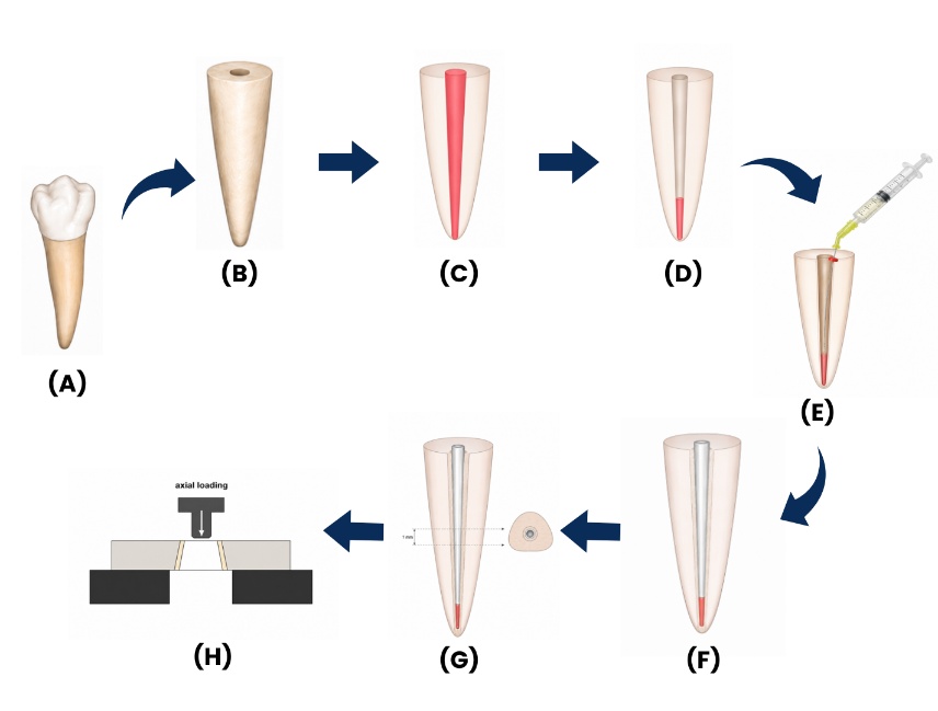
Each root was sectioned perpendicular to its long axis using a precision cutting machine to obtain standardized 2-mm thick slices from the middle third of the root.
Push-out bond strength testing was performed using a universal testing machine. Each specimen was positioned with the apical surface facing upward, and a compressive load was applied using a cylindrical plunger at a crosshead speed of 0.5 mm/min until dislodgement of the fiber post occurred.
The maximum load required for post extrusion was recorded in Newtons (N), and push-out bond strength was calculated in megapascals (MPa) using the formula:
Push-out bond strength (MPa) = Maximum load (N) / Bonded surface area (mm²)
A schematic overview of the specimen preparation and push-out bond strength testing procedure is presented in Figure 1.
2.10. Statistical analysis
Statistical analysis was performed using SPSS software version 21.0 (IBM Corp., NY, USA).
The mean and standard deviation values were calculated for each group.
Intergroup comparison of push-out bond strength values among the four groups was performed using one-way analysis of variance (ANOVA) followed by Tukey’s post hoc test.
A p-value < 0.05 was considered statistically significant.
3. Results
Statistical analysis was performed using SPSS software version 21.0 (IBM Corp., NY, USA). Descriptive statistics including mean and standard deviation were calculated for all groups. Intergroup comparison of push-out bond strength values among the four experimental groups was performed using one-way ANOVA, followed by Tukey’s post hoc test. A p-value < 0.05 was considered statistically significant.
|
Group |
Dentin Pretreatment Agent |
Mean Push-Out Bond Strength (MPa) |
|---|---|---|
|
Group I |
1-ethyl-3-(3-dimethylaminopropyl) carbodiimide (EDC) |
12.8 ± 1.9 |
|
Group II |
Proanthocyanidin (PAC) |
11.8 ± 1.6 |
|
Group III |
0.2% Chitosan |
9.8 ± 1.5 |
|
Group IV |
Saline (Control) |
6.0 ± 1.2 |
The mean push-out bond strength values demonstrated statistically significant differences among the groups (p < 0.001). The highest push-out bond strength was observed in Group I (EDC) with a mean value of 12.8 ± 1.9 MPa, followed by Group II (Proanthocyanidin) (11.8 ± 1.6 MPa), Group III (0.2% Chitosan) (9.8 ± 1.5 MPa), and Group IV (Saline control) (6.0 ± 1.2 MPa) as presented in Table 1.
Tukey’s post hoc analysis revealed that Group I (EDC) and Group II (Proanthocyanidin) exhibited comparable push-out bond strength values with no statistically significant difference between them (p > 0.05). Both groups demonstrated significantly higher bond strength compared with Group III (Chitosan) and Group IV (Saline control) (p < 0.05).
Group III (0.2% Chitosan) demonstrated significantly higher push-out bond strength compared with the saline control group (p < 0.05), indicating improved interaction between the modified dentin substrate and resin cement. However, chitosan showed lower bond strength values compared with EDC and proanthocyanidin groups.
Overall, dentin pretreatment with collagen-modifying agents enhanced fiber post retention, with EDC and proanthocyanidin showing the greatest improvement in push-out bond strength, followed by chitosan, whereas saline-treated specimens demonstrated the lowest values.
4. Discussion
The long-term success of fiber post restorations depends largely on the integrity and durability of the bond between the resin cement and radicular dentin. Although fiber posts exhibit favorable mechanical properties and stress distribution similar to dentin, debonding at the resin cement–dentin interface remains one of the major causes of clinical failure.[3] The present in vitro study evaluated the effect of different dentin pretreatment agents, including EDC, proanthocyanidin, and chitosan, on the push-out bond strength of fiber posts luted with resin cement.
The push-out test was selected for evaluating bond strength because it provides a reliable assessment of the adhesion between the post and surrounding dentin by generating stress distribution similar to clinical loading conditions.[11] In the present study, a single type of fiber post system and resin luting cement were used for all specimens to minimize variations related to restorative materials and ensure standardization of the bonding protocol. This allowed the differences observed in push-out bond strength values to be primarily associated with the effect of the dentin pretreatment agents. The middle third of the root was selected for evaluation as it provides a relatively uniform post space geometry and minimizes anatomical variations associated with the coronal and apical regions.[12]
In the present study, dentin pretreatment with all biomodifying agents resulted in higher push-out bond strength values compared with saline-treated specimens. The increase in bond strength observed with EDC, proanthocyanidin, and chitosan may be attributed to their ability to modify the dentinal collagen matrix, improve substrate stability, and enhance interaction between resin cement and root dentin.
Group I (EDC) demonstrated the highest push-out bond strength (12.8 ± 1.9 MPa) among all groups. The improved bonding performance of EDC may be attributed to its collagen cross-linking ability. EDC acts as a zero-length cross-linking agent by activating carboxyl groups present within collagen molecules, resulting in the formation of stable amide bonds between adjacent collagen chains. This cross-linking mechanism increases collagen stiffness, reduces susceptibility to enzymatic degradation, and improves the mechanical properties of dentin.[5] However, despite its superior bonding performance, the synthetic nature of EDC has stimulated the search for naturally derived dentin biomodifiers. Moreover, the efficacy of EDC is concentration- and application time-dependent, with variations in these parameters influencing the extent of collagen stabilization and its interaction with dentinal tissues.[4] Therefore, naturally derived agents such as proanthocyanidins and chitosan have gained attention due to their collagen-stabilizing properties, antioxidant potential, and favorable biocompatibility, offering promising alternatives for enhancing dentin bonding durability.
Group II (Proanthocyanidin) demonstrated comparable push-out bond strength values (11.8 ± 1.6 MPa) to EDC, with no statistically significant difference observed between the two groups. The enhanced bonding ability of grape seed-derived proanthocyanidin (GSE-PAC) may be attributed to its strong interaction with dentinal collagen fibrils. PACs are oligomeric flavonoids containing multiple hydroxyl groups that facilitate hydrogen bonding and hydrophobic interactions with collagen, resulting in increased intermolecular cross-linking within the dentin matrix.[13] This biomodification improves collagen rigidity, enhances resistance against enzymatic degradation, and provides a more stable substrate for resin cement interaction. However, studies have shown that the clinical application of GSE-PAC may be influenced by its concentration-dependent activity and variability in composition, as the quantity and structure of proanthocyanidins can differ according to extraction methods and natural sources.[14] These limitations have encouraged the exploration of other naturally derived biomodifiers with additional functional properties.
Group III (0.2% Chitosan) demonstrated improved push-out bond strength (9.8 ± 1.5 MPa) compared with the saline control group, although the values were lower than those observed in the EDC and proanthocyanidin groups. The favorable effect of chitosan may be attributed to its unique physicochemical properties, including biocompatibility, antimicrobial activity, and interaction with dentinal components. The protonated amino groups present in chitosan molecules can interact electrostatically with negatively charged dentinal structures and may contribute to modification of the collagen matrix. Additionally, hydrogen bonding and intermolecular interactions between chitosan and dentinal collagen may enhance collagen stability and improve the interaction between resin cement and radicular dentin.[15] However, the lower bond strength compared with EDC and proanthocyanidin may be related to its comparatively weaker collagen cross-linking potential. Further investigations evaluating chitosan in nanoparticulate form may be warranted, as chitosan nanoparticles offer increased surface area and may enhance interaction with dentinal collagen networks and substrate penetration, potentially improving dentin biomodification efficacy and long-term bond durability.
The saline control group exhibited the lowest push-out bond strength (6.0 ± 1.2 MPa), representing the baseline adhesion achieved without dentin biomodification. In untreated dentin, exposed collagen fibrils remain more susceptible to enzymatic degradation by endogenous proteolytic enzymes such as matrix metalloproteinases (MMPs) and cysteine cathepsins, which may compromise the stability of the resin–dentin interface.[2][4] The significantly higher bond strength observed in biomodifier-treated groups highlights the importance of dentin substrate modification before fiber post cementation.
The present study has certain limitations. Being an in vitro investigation, it does not completely replicate intraoral conditions such as thermal cycling, mechanical fatigue, and long-term enzymatic degradation. Further studies incorporating aging protocols, cyclic loading, and evaluation of long-term bond durability are required to validate the clinical applicability of these dentin biomodifying agents.
Overall, the findings indicate that pretreatment of radicular dentin with collagen-modifying agents significantly enhances fiber post retention. Among the tested materials, EDC and proanthocyanidin demonstrated the greatest improvement in push-out bond strength, whereas chitosan showed promising potential as a natural biomaterial-based pretreatment agent.
5. Conclusion
Dentin pretreatment with collagen-modifying agents significantly improved the push-out bond strength of fiber posts compared with untreated group. EDC and proanthocyanidin demonstrated comparable and higher bond strength values, while 0.2% chitosan showed improved adhesion compared with untreated dentin. These findings suggest that dentin biomodification may enhance fiber post retention and improve the resin cement–dentin interface.
6. Authors Contributions
Tanvi Pimpale: Conceptualization, Data curation, Formal analysis, Funding acquisition, Investigation, Methodology, Validation, Writing – original draft, Writing – review editing, Gaurav Jain: Conceptualization, Data curation, Formal analysis, Funding acquisition, Investigation, Methodology, Project administration, Resources, Software, Supervision, Validation, Visualization, Writing – original draft, Writing – review editing, Sonali Verma: Conceptualization, Data curation, Formal analysis, Funding acquisition, Project administration, Writing – original draft, Writing – review editing, Pradyumna Misra: Conceptualization, Project administration, Supervision.
7. Source of Funding
None
8. Conflicts of Interest
None
References
- Dietschi D, Duc O, Krejci I, Sadan A. Biomechanical considerations for the restoration of endodontically treated teeth: a systematic review of the literature--Part 1. Composition and micro- and macrostructure alterations. Quintessence Int. 2007;38(9):733-43. [Google Scholar]
- Mannocci F, Cowie J. Restoration of endodontically treated teeth. Br Dent J. 2014;216(6):341-6. [Google Scholar] [Crossref]
- Nivaskumar GA, Ranjini MA, Vedavathi B, Ambili C, Prathiksha SK. Effect of resveratrol pretreatment of radicular dentin on immediate and delayed pushout bond strength of fiber post luted using self-adhesive resin cement: An in vitro study. J Conserv Dent Endod. 2023;26(5):579-83. [Google Scholar] [Crossref]
- Schestakow A, Huang CM, Yin IX, Chu CH. The role of collagen cross-linker in dentin mineralization: a scoping review. J Dent. 2025;158:105799. [Google Scholar] [Crossref]
- Mazzoni A, Apolonio FM, Saboia VP, Santi S, Angeloni V, Checchi V. Carbodiimide inactivation of MMPs and effect on dentin bonding. J Dent Res. 2014;93(3):263-8. [Google Scholar] [Crossref]
- Cadenaro M, Fontanive L, Navarra CO, Gobbi P, Mazzoni A, Di Lenarda R. Effect of carboidiimide on thermal denaturation temperature of dentin collagen. Dent Mater. 2016;32(4):492-8. [Google Scholar] [Crossref]
- Castellan CS, Pereira PN, Grande RH, Bedran-Russo AK. Mechanical characterization of proanthocyanidin-dentin matrix interaction. Dent Mater. 2010;26(10):968-73. [Google Scholar] [Crossref]
- Green B, Yao X, Ganguly A, Xu C, Dusevich V, Walker MP. Grape seed proanthocyanidins increase collagen biodegradation resistance in the dentin/adhesive interface when included in an adhesive. J Dent. 2010;38(11):908-15. [Google Scholar] [Crossref]
- Silva PV, Guedes DF, Nakadi FV PJCFA. Chitosan: a new solution for removal of smear layer after root canal instrumentation. Int Endod J. 2013;46(4):332-8. [Google Scholar] [Crossref]
- Paschoini VL, Ziotti IR, Neri CR, Corona SAM, Souza-Gabriel AE. Chitosan improves the durability of resin-dentin interface with etch-and-rinse or self-etch adhesive systems. J Appl Oral Sci. 2021;29:e20210356. [Google Scholar] [Crossref]
- Goracci C, Tavares AU, Fabianelli A, Monticelli F, Raffaelli O, Cardoso PC. The adhesion between fiber posts and root canal walls: comparison between microtensile and push-out bond strength measurements. Eur J Oral Sci. 2004;112(4):353-61. [Google Scholar] [Crossref]
- Soares CJ, Santana FR, Castro CG, Santos-Filho PC, Soares PV, Qian F. Finite element analysis and bond strength of a glass post to intraradicular dentin: comparison between microtensile and push-out tests. Dent Mater. 2008;24(10):1405-11. [Google Scholar] [Crossref]
- Vidal CM, Zhu W, Manohar S, Aydin B, Keiderling TA, Messersmith PB. Collagen-collagen interactions mediated by plant-derived proanthocyanidins: A spectroscopic and atomic force microscopy study. Acta Biomater. 2016;41:110-8. [Google Scholar] [Crossref]
- Enrich-Essvein T GLSRNACJC, Maravic T, Mazzitelli C. Effects of proanthocyanidin-functionalized hydroxyapatite nanoparticles on dentin bonding. Clin Oral Investig. 2024;28(8):444. [Google Scholar] [Crossref]
- Ziotti IR, Paschoini VL, Corona SAM, Souza-Gabriel AE. Chitosan-induced biomodification on demineralized dentin to improve the adhesive interface. Restor Dent Endod. 2022;47(3):e28. [Google Scholar] [Crossref]
- Abstract
- 1. Introduction
- 2. Materials and Methods
- 2.1. Study design
- 2.2. Sample selection and preparation
- 2.3. Root canal treatment
- 2.4. Post space preparation
- 2.5. Preparation of dentin pretreatment solutions
- 2.5.1. Preparation of 0.5 M 1-Ethyl-3-(3-dimethylaminopropyl) carbodiimide hydrochloride (EDC) solution
- 2.5.2. Preparation of 6.5% proanthocyanidin solution
- 2.5.3. Preparation of 0.2% chitosan solution
- 2.6. Group allocation
- 2.7. Dentin pretreatment protocol
- 2.8. Fiber post cementation
- 2.9. Push-out bond strength evaluation
- 2.10. Statistical analysis
- 3. Results
- 4. Discussion
- 5. Conclusion
- 6. Authors Contributions
- 7. Source of Funding
- 8. Conflicts of Interest
- References
Article Metrics
- Visibility 287 Views
- Downloads 337 Views
- DOI 10.18231/j.jicd.57125.1785131551
-
CrossMark

- Citation
- Received Date June 04, 2026
- Accepted Date July 10, 2026
- Publication Date July 27, 2026