Journal of International College of Dentists
Journal of International College of Dentists (JICD) is an open-access, peer-reviewed, Quarterly journal publishing under the auspices of the International College of Dentists. As soon as the Article is accepted for Publication, it will be placed in “Ahead of Print” on the journal page with the aim of rapid and better knowledge dissemination. In addition, the journal allows free access (Open Access) to its contents, which is likely to attract more readers and citations to articles published in journal. Manuscripts must be prepared under the “Uniform requirements” of the ...
Association of anterior and posterior occlusal planes with sella-nasion and Frankfort horizontal planes in different skeletal malocclusion- a cephalometric study
Abstract
Introduction: The occlusal plane plays an important role in craniofacial morphology and is associated with different skeletal malocclusion patterns. Class II malocclusions are commonly associated with steeper occlusal planes, whereas Class III malocclusions exhibit flatter occlusal planes. To better represent the sagittal curvature of occlusion, the occlusal plane can be divided into anterior and posterior occlusal planes.
Aim and Objectives: To evaluate the relationship of the anterior occlusal plane (AOP) and posterior occlusal plane (POP) with the Sella-Nasion (SN) plane and Frankfort Horizontal (FH) plane in different skeletal malocclusion patterns.
Materials and Methods: Pretreatment lateral cephalograms of 60 subjects were divided into three groups: Skeletal Class I (ANB = 0°–4°), Skeletal Class II (ANB > 4°), and Skeletal Class III (ANB < 0°), with 20 subjects in each group. Angular measurements evaluated were SN-POP, FH-POP, SN-AOP, and FH-AOP. Statistical analysis was performed using one-way ANOVA followed by Post Hoc analysis.
Results: Statistically significant differences were observed for SN-POP (p = 0.003), FH-POP (p = 0.016), and SN-AOP (p = 0.009). Class II subjects exhibited steeper anterior and posterior occlusal plane inclinations compared with Class I and Class III subjects. FH-AOP did not show a statistically significant difference among the groups (p = 0.103).
Conclusion: Skeletal malocclusion patterns influence anterior and posterior occlusal plane inclination. Class II malocclusion demonstrated steeper SN-POP, FH-POP, and SN-AOP angles, whereas Class III malocclusion exhibited flatter occlusal plane inclinations, indicating a close association between occlusal plane orientation and skeletal morphology.
1. Introduction
The occlusal plane is an important component of the craniofacial complex and plays a significant role in dental esthetics, masticatory function, and orthodontic diagnosis and treatment planning. Its orientation influences the sagittal and vertical relationships of the jaws and has been shown to be associated with different skeletal malocclusion patterns. Alterations in the inclination of the occlusal plane may affect mandibular position, facial profile, and overall dentofacial harmony.[1][2][3][4]
Several studies have reported a relationship between occlusal plane inclination and skeletal morphology. A steeper occlusal plane is commonly associated with skeletal Class II malocclusion, whereas a flatter occlusal plane is often observed in skeletal Class III malocclusion. These variations may reflect differences in maxillomandibular growth patterns and dentoalveolar compensation mechanisms.[5][6][7]
Traditionally, the occlusal plane has been represented as a single straight line in cephalometric analyses. However, this approach may not adequately represent the natural curvature of the dental arches. To better describe the sagittal characteristics of occlusion, Fushima et al. subdivided the maxillary occlusal plane into anterior occlusal plane (AOP) and posterior occlusal plane (POP) at the level of the maxillary second premolar. This subdivision provides a more detailed assessment of occlusal plane inclination and its relationship with craniofacial structures.[5]
The Sella-Nasion (SN) plane and Frankfort Horizontal (FH) plane are widely used cranial reference planes in cephalometric analysis. Evaluating the relationship of anterior and posterior occlusal planes with these reference planes may provide valuable information regarding skeletal discrepancies and occlusal characteristics associated with different malocclusion patterns.[1][2][8]
Although the association between occlusal plane inclination and skeletal malocclusion has been investigated previously, limited studies have evaluated both anterior and posterior occlusal planes in relation to the SN and FH planes across different skeletal malocclusion groups. Therefore, the present study was undertaken to evaluate the association of anterior and posterior occlusal planes with the SN and FH planes in skeletal Class I, Class II, and Class III malocclusions.
2. Materials and Methods
2.1. Study design and sample selection
This retrospective cephalometric study was conducted in the Department of Orthodontics and Dentofacial Orthopaedics at National Dental College and Hospital. Pretreatment lateral cephalograms of 60 subjects were selected from the departmental records.
The subjects were divided into three groups based on their skeletal relationship as determined by the ANB angle: Group I (n = 20): Skeletal Class I (ANB = 0°–4°). Group II (n = 20): Skeletal Class II (ANB > 4°). Group III (n = 20): Skeletal Class III (ANB < 0°).
2.2. Inclusion criteria
The following criteria were used for sample selection: Presence of permanent dentition excluding third molars. Availability of good-quality pretreatment lateral cephalograms.
2.3. Exclusion criteria
Subjects presenting with any of the following conditions were excluded from the study: Missing teeth. Craniofacial syndromes such as cleft lip and palate. Previous orthodontic treatment. Temporomandibular joint abnormalities. Poor-quality or distorted cephalograms.
2.4. Cephalometric analysis
All lateral cephalograms were traced and analysed for the identification of skeletal and dental landmarks. The following reference planes were used: SN plane: Line joining Sella (S) and Nasion (N). FH plane: Line joining Porion (Po) and Orbitale (Or). PP (Palatal plane): Line joining Anterior Nasal Spine (ANS) and Posterior Nasal Spine (PNS). AOP (Anterior occlusal plane): Line joining the maxillary incisal edge and the cusp tip of the maxillary second premolar. POP (Posterior occlusal plane): Line joining the cusp tip of the maxillary second premolar and the midpoint between the cusp tips of the maxillary second molar. MP (Mandibular plane): Line joining Tangential Gonion and Menton.
The following angular measurements were evaluated: SN-AOP: Angle between the SN plane and anterior occlusal plane. SN-POP: Angle between the SN plane and posterior occlusal plane. FH-AOP: Angle between the FH plane and anterior occlusal plane. FH-POP: Angle between the FH plane and posterior occlusal plane. ANB: Angle formed by points A, Nasion, and B.
3. Statistical Analysis
The collected data were entered into Microsoft Excel and analysed statistically. Descriptive statistics including mean and standard deviation were calculated for all variables. Intergroup comparisons among skeletal Class I, Class II, and Class III malocclusion groups were performed using One-Way Analysis of Variance (ANOVA). Post Hoc analysis was carried out to identify pairwise differences between the groups whenever a statistically significant difference was observed. A p-value of less than 0.05 was considered statistically significant.
4. Results
The mean and standard deviation values of the angular measurements for the three skeletal malocclusion groups are presented in Table 1.
The angular measurements of anterior and posterior occlusal planes relative to the SN and FH planes were compared among skeletal Class I, Class II, and Class III malocclusions and illustrated in Graph 1.
The SN-POP angle demonstrated statistically significant differences among the three skeletal malocclusion groups (p = 0.003). The mean SN-POP angle was highest in the Class II group (22.850 ± 6.450°), followed by the Class I group (17.150 ± 6.072°) and the Class III group (16.400 ± 5.906°). Post Hoc analysis revealed significant differences between Class I and Class II malocclusions and between Class II and Class III malocclusions. ([Table 1]), (Graph 1)
Similarly, the FH-POP angle showed a statistically significant difference among the groups (p = 0.016). The Class II group exhibited the highest mean value (15.000 ± 5.525°), whereas the Class I and Class III groups showed comparable values of 10.600 ± 5.393° and 10.675 ± 5.151°, respectively. Post Hoc analysis demonstrated significant differences between Class I and Class II malocclusions and between Class II and Class III malocclusions. ([Table 1]), (Graph 1)
The SN-AOP angle also demonstrated a statistically significant difference among the three groups (p = 0.009). The highest mean value was observed in the Class II group (17.700 ± 6.489°), followed by the Class I group (14.450 ± 6.916°), while the Class III group exhibited the lowest mean value (10.800 ± 7.015°). Post Hoc analysis revealed a significant difference between Class II and Class III malocclusions. ([Table 1]), (Graph 1)
|
Angle |
Malocclusion |
Mean ± Std Dev |
p-value |
Post hoc analysis |
|---|---|---|---|---|
|
SN-POP |
Class I |
17.150 ± 6.072 |
0.003 |
Significant difference between Class I and Class II, Class II and III |
|
Class II |
22.850 ± 6.450 |
|||
|
Class III |
16.400 ± 5.906 |
|||
|
FH-POP |
Class I |
10.600 ± 5.393 |
0.016 |
Significant difference between Class I and Class II, Class II and III |
|
Class II |
15.000 ± 5.525 |
|||
|
Class III |
10.675 ± 5.151 |
|||
|
SN-AOP |
Class I |
14.450 ± 6.916 |
0.009 |
Significant difference between Class II and III |
|
Class II |
17.700 ± 6.489 |
|||
|
Class III |
10.800 ± 7.015 |
|||
|
FH-AOP |
Class I |
7.850 ± 5.460 |
0.103 |
No statistically significant difference |
|
Class II |
9.550 ± 7.022 |
|||
|
Class III |
4.800 ± 8.230 |
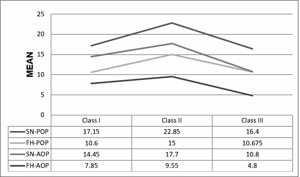
In contrast, the FH-AOP angle did not demonstrate a statistically significant difference among the skeletal malocclusion groups (p = 0.103). The mean values were 7.850 ± 5.460° for Class I, 9.550 ± 7.022° for Class II, and 4.800 ± 8.230° for Class III malocclusions. ([Table 1]), (Graph 1)
Overall, Class II malocclusion demonstrated steeper anterior and posterior occlusal plane inclinations relative to both SN and FH reference planes, whereas Class III malocclusion exhibited comparatively flatter occlusal plane inclinations.
5. Discussion
The occlusal plane plays a fundamental role in craniofacial morphology and has long been considered an important parameter in orthodontic diagnosis and treatment planning. Variations in occlusal plane inclination have been associated with different skeletal malocclusion patterns and may influence both facial aesthetics and functional occlusion.
To better evaluate these relationships, the present study assessed the association of anterior and posterior occlusal planes with the Sella-Nasion (SN) and Frankfort Horizontal (FH) planes in subjects with skeletal Class I, Class II, and Class III malocclusions.
As shown in [Table 1] and Graph 1, significant differences were observed in SN-POP, FH-POP, and SN-AOP among the skeletal malocclusion groups. The posterior occlusal plane measurements demonstrated the most pronounced differences, particularly in skeletal Class II subjects. The mean SN-POP angle was significantly greater in Class II individuals than in Class I and Class III subjects. Similarly, the FH-POP angle was significantly increased in Class II malocclusion. These findings indicate a steeper posterior occlusal plane in Class II skeletal patterns. Similar findings were reported by Fushima et al. and Tanaka and Sato, who observed that the posterior occlusal plane tends to be steeper in skeletal Class II malocclusions and flatter in skeletal Class III malocclusions.[5][6]
The increased inclination of the posterior occlusal plane observed in Class II malocclusion may be related to the characteristic craniofacial morphology of these patients. Skeletal Class II individuals frequently exhibit mandibular retrusion and a tendency toward backward mandibular rotation, resulting in a steeper occlusal plane orientation.[9][10][11] In contrast, Class III subjects demonstrated comparatively flatter posterior occlusal plane inclinations, which may be associated with forward mandibular positioning and a more horizontal growth pattern.[7][12] Ardani et al. also reported significant differences in occlusal plane inclination between skeletal Class I and Class III malocclusions and suggested that occlusal plane orientation may serve as a useful diagnostic indicator for skeletal Class III discrepancies.[13] Similarly, Sato emphasized the importance of posterior occlusal plane inclination in the development and characterization of skeletal Class III malocclusion, highlighting its close relationship with craniofacial growth patterns.[12] Reyes et al., reported greater maxillary molar eruption in skeletal Class III subjects compared with individuals with normal occlusion, a finding that may contribute to the flatter posterior occlusal plane observed in Class III malocclusion.[14]
The anterior occlusal plane also demonstrated significant variation among the skeletal groups. The SN-AOP angle was highest in Class II subjects and lowest in Class III subjects, indicating that the anterior segment of the occlusal plane follows a similar pattern to that observed for the posterior segment. As illustrated in Graph 1, Class II malocclusion consistently exhibited greater angular values, whereas Class III malocclusion demonstrated comparatively flatter occlusal plane inclinations.
Although the FH-AOP angle showed differences among the groups, the overall comparison did not reach statistical significance. However, a noticeable difference was observed between Class II and Class III subjects. The absence of statistical significance may be attributed to limitations associated with the Frankfort Horizontal plane. The FH plane is dependent on the identification of porion and orbitale, landmarks that may exhibit considerable individual variation and are often more difficult to locate precisely on lateral cephalograms. Consequently, measurements based on the SN plane may provide a more reliable assessment of occlusal plane inclination. Similar observations were reported by Čelar et al., who found that anterior occlusal plane inclinations did not differ significantly among skeletal malocclusion groups, whereas posterior occlusal plane inclinations showed significant differences.[15]
The findings of the present study are consistent with previous investigations that have reported steeper occlusal planes in skeletal Class II malocclusions and flatter occlusal planes in skeletal Class III malocclusions.[12] The subdivision of the occlusal plane into anterior and posterior components allows a more detailed assessment of sagittal occlusal relationships and may provide additional diagnostic information beyond conventional occlusal plane measurements.[5][6][15]
The graphical representation in Graph 1 further highlights the distinct pattern of occlusal plane inclination among the skeletal groups. The consistently higher SN-POP, FH-POP, and SN-AOP values in Class II subjects and lower values in Class III subjects emphasize the influence of skeletal pattern on occlusal plane orientation.
5.1. Limitations of the study
The present study was limited by a relatively small sample size and its retrospective design. Furthermore, only angular cephalometric measurements were evaluated, and soft tissues as well as functional parameters were not considered. Future studies involving larger samples and three-dimensional imaging techniques may provide a more comprehensive understanding of the relationship between occlusal plane inclination and skeletal malocclusion.
6. Conclusion
The study provides important cephalometric data on the relationship between the anterior and posterior occlusal planes and the Sella-Nasion and Frankfort Horizontal planes across different skeletal malocclusions.
Statistically significant differences were observed for most of the measured angles i.e. SN-POP, FH-POP, SN-AOP and statistically non-significant difference for FH-AOP, highlighting the influence of skeletal class on craniofacial morphology.
The study concluded that Class II malocclusion showed steeper SN-POP, FH-POP, SN-AOP angle and Class III malocclusion showed flatter SN-POP, FH-POP, SN-AOP angle whereas Class I malocclusion showed flatter SN-POP, FH-POP angle.
Further studies with larger sample sizes would help solidify these findings.
7. Authors Contributions
Megha Girdhar: Conceptualization, data curation, formal analysis, investigation, methodology, resources, software, scientific advisor, visualization, writing – original draft, writing – review editing. Harvinder Singh: Conceptualization, data curation, formal analysis, investigation, methodology, resources, software, scientific advisor, visualization. Vinay Dua: Conceptualization, data curation, formal analysis, software, scientific advisor, visualization. Yenika Manchanda: Conceptualization, investigation, methodology, resources, software, scientific advisor, visualization. Amanjot Kaur: Conceptualization, data curation, formal analysis, investigation, methodology, scientific advisor.
8. Source of Funding
None.
9. Conflict of Interest
None.
References
- Downs WB. Variations in facial relationships; their significance in treatment and prognosis. Am J Orthod. 1948;34(10):812-840. [Google Scholar] [Crossref]
- Sassouni V. A classification of skeletal facial types. Am J Orthod. 1969;55(2):109-123. [Google Scholar] [Crossref]
- Proffit WR, Fields H, Larson B, Sarver DM. Contemporary Orthodontics. 6th ed. St Louis: Elsevier; 2019. [Google Scholar] https://shop.elsevier.com/books/contemporary-orthodontics/proffit/978-0-323-54387-3
- Nissan J, Barnea E, Zeltzer C, Cardash HS. Relationship between occlusal plane determinants and craniofacial structures. J Oral Rehabil. 2003;30(6):587-591. [Google Scholar] [Crossref]
- Fushima K, Kitamura Y, Mita H, Sato S, Suzuki Y, Kim YH. Significance of the cant of the posterior occlusal plane in class II division 1 malocclusions. Eur J Orthod. 1996;18(1):27-40. [Google Scholar] [Crossref]
- Tanaka EM, Sato S. Longitudinal alteration of the occlusal plane and development of different dentoskeletal frames during growth. Am J Orthod Dentofacial Orthop. 2008;134(5):602-602.e11. e1–11; discussion 602–3. [Google Scholar] [Crossref]
- Alhammadi MS. Dentoalveolar compensation in different anterioposterior and vertical skeletal malocclusions. J Clin Exp Dent. 2019;11(8):e745-e753. [Google Scholar] [Crossref]
- Kim YH, Vietas JJ. Anteroposterior dysplasia indicator: an adjunct to cephalometric differential diagnosis. Am J Orthod. 1978;73(6):619-633. [Google Scholar] [Crossref]
- Schudy FF. The rotation of the mandible resulting from growth: Its implications in orthodontic treatment. Angle Orthod. 1965;35:36-50. [Google Scholar] [Crossref]
- Nanda SK. Growth patterns in subjects with long and short faces. Am J Orthod Dentofacial Orthop. 1990;98(3):247-258. [Google Scholar] [Crossref]
- Bishara SE, Jakobsen JR. Longitudinal changes in three normal facial types. Am J Orthod. 1985;88(6):466-502. [Google Scholar] [Crossref]
- Sato S. Case report: developmental characterization of skeletal Class III malocclusion. Angle Orthod. 1994;64(2):105-111. discussion 111–2. [Google Scholar] [Crossref]
- Ardani IGAW, Wicaksono A, Hamid T. The Occlusal Plane Inclination Analysis for Determining Skeletal Class III Malocclusion Diagnosis. Clin Cosmet Investig Dent. 2020;12:163-171. [Google Scholar] [Crossref]
- Reyes BC, Baccetti T, McNamara JA Jr. An estimate of craniofacial growth in Class III malocclusion. Angle Orthod. 2006;76(4):577-584. [Google Scholar] [Crossref]
- Čelar A, Tafaj E, Graf A, Lettner S. Association of anterior and posterior occlusal planes with different Angle and skeletal classes in permanent dentitions: A lateral cephalometric radiograph study. J Orofac Orthop. 2018;79(4):267-276. [Google Scholar] [Crossref]
Article Metrics
- Visibility 121 Views
- Downloads 152 Views
- DOI 10.18231/j.jicd.55364.1792217764
-
CrossMark

- Citation
- Received Date June 25, 2026
- Accepted Date July 29, 2026
- Publication Date October 17, 2026